Katie Meyers
Unit Three Lab Project: Build a Movable Limb
This is my model of a movable limb and the cellular activity responsible for muscle contraction. I decided to use the biceps brachii muscle for the main muscle in this lab. I also have a working elbow hinge joint. There is a shoulder ball-and-socket joint present in the pictures, but it is there only to complete the model and make the upper connection of the biceps brachii muscle to the scapula possible. So the reader is aware, my captions are above the pictures they pertain to.
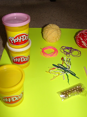
Here are several images of the objects I used to make my model(s). The items are as follows: leopard glove, two different types of furniture wheels, PVC pipe, unsharpened pencil, blue straw, red streamer paper, duct tape, Knex, putty, seven colors of play dough (pink, white, yellow, light blue, red, orange, darker blue), plastic string (pink and purple), yarn (red, white, yellow), multi-colored twist ties, tiny jingle bells, red and white striped straws, rope, lime green posterboard. I will specify what each item was used for later in this presentation, in the captions of each of my detailed model pictures.


I began making my limb by putting together the humerus (PVC pipe), elbow hinge joint (brown hinge furniture wheel), shoulder ball-and-socket joint (brass ball-and-socket furniture wheel), and the points of the scapula to which the bicep muscle will attach to (two yellow Knex and a green Knex connector [covered in duct tape]). I put it all together with trusty duct tape.

Here is a labeled and finished image of my arm, with the bicep brachii muscle in a relaxed (straighter) position.

I used a leopard skin pattern glove to represent the hand, a thick non-sharpened decorated pencil for the ulna bone, and a blue straw for the radius bone. These were attached to the elbow joint with putty.

This is a close-up of the aforementioned elbow hinge joint, also showing the bicep brachii (red streamer paper) attached to the radius bone.

Here is a close-up of my PVC pipe humerus and red streamer paper relaxed biceps brachii.

Here is an image of the top of the humerus attached to the aforementioned ball-and-socket shoulder joint.

Here is an image of the two places where the biceps brachii attaches to the scapula (Knex), which is attached to the shoulder joint with duct tape.

Here is a picture of an axon. The axon is made of rope, the axon terminal happens to be the frayed end of that rope, and the Schwann cells are made of duct tape. The parts of the axon exposed between the Schwann cells are called nodes of Ranvier.

The axon carries action potentials, or messages that are sent to signal muscle contraction.

Here is a close-up of the three stages of action potential and how it works in the axon. The first stage is resting potential, which is the condition in which the axon remains between action potentials. Notice there are more potassium than sodium ions inside the axon, and more sodium than potassium ions outside the axon. In this stage, the membrane potential is -65. The sodium ions and gates are made from light blue play dough, and the potassium ions and gates are made from pink play dough.

This is an image of the beginning step of an action potential. The sodium gates open, while the potassium gates remain closed. This results in more ions inside the axon than outside the axon and a membrane potential of +40.

Here is an image of the ending step of an action potential. The sodium gates close, while the potassium gates open. This evens out the number of ions inside and outside the axon, thereby returning the membrane potential to -65.

After this action potential (red yarn) travels down an axon (dark blue play dough), it comes to a muscle cell. It then goes through the neuromuscular junction (thick chunk of dark blue play dough), travels across the sarcolemma (orange play dough), and goes into the muscle cell by way of T tubules (red and white striped straws), which extend through the sarcoplasmic reticulum (white play dough) and into the myofibril (yellow play dough).

Here is a close-up introducing how action potentials, sarcoplasmic reticulum (white play dough), T tubules (red and white striped straw), and calcium (pink play dough) interact. When an action potential is absent, the sarcoplasmic reticulum stores calcium. The red play dough is just a background device to make the model parts easier to see.

However, when an action potential (red yarn) travels down the T tubule, it stimulates the sarcoplasmic reticulum to release calcium into the myofibril (background lime green posterboard), where it will then be used for actin and myosin reactions.

How does calcium interact with actin and myosin? Well, to start with, here is an actin strand (or filament) (tiny jingle bells on purple plastic string) covered in its usual troponin (light blue play dough) and tropomyosin (yellow yarn) proteins. However, you will also see that the released calcium (now white play dough) has attached itself to the troponin.

This calcium attachment causes a reaction that pulls the tropomyosin away from the actin filament, thereby exposing the myosin binding sites (“X”s on jingle bells), making interaction between actin and myosin possible.

Here is an example of a sarcomere, which is the name of the overall setup of actin (tiny jingle bells strung on purple and pink plastic string) and myosin (red play dough around many multi-colored twist ties) in muscle cells. The myosin has cross-bridges (multi-colored twist ties) on it, thereby enabling it to perform the next step in muscle contraction. This is an image of a relaxed sarcomere, before the abovementioned calcium reaction has made actin and myosin interaction possible.

This is a diagram of how a contracted sarcomere looks. The sarcomere contracts after the calcium has made actin-myosin binding possible, and the myosin has used its cross-bridges to pull actin together.

Finally, after all these steps occurring on a microscopic level and in many muscle cells, our muscle, the biceps brachii, contracts! This muscle bends the elbow hinge joint, pulling the forearm closer to the upper arm.

Here is a close-up of the contracted muscle bending the elbow joint.

Through these models, pictures, and captions, I have attempted to show and describe on a molecular and cellular level how muscles move. I began by showing a relaxed muscle, the biceps brachii, in the body and how it is attached in the human body. Then, I delved into the molecular steps, beginning with an action potential traveling down an axon and ending with the reactions of actin and myosin filaments. As a result of all these microscopic movements and reactions in the many muscle cells that together form the biceps brachii, this muscle contracts. The result of a contracting biceps brachii muscle is that the elbow joint bends and moves the forearm in a flexion direction (meaning the joint angle decreases).
Although this project was just as large and difficult to tackle as the cell project, I learned just as much about the particular subject as I did then, if not more. By doing this assignment, all the information in this unit about movement has finally sunk in. At first, these concepts about movement beginning at the microscopic level were hard to remember and put together. However, by doing this project in a step-by-step fashion of how the whole machine works together, I finally realized the sequencing of events and how they affected each other. I learned a lot from this project, making it very worthwhile. It was a long, hard road, but worthwhile in the end.

No comments:
Post a Comment