Thursday, July 12, 2007

Compendium Six – Body Movement: Muscles, Joints, and Bones

Katie Meyers

Compendium Six – Body Movement: Muscles, Joints, and Bones

Muscles

  • Muscle names
  • Muscle movement overview
  • Muscle tissue
    • 3 types
  • Muscle fiber cell
  • Motor unit
  • Muscle’s 4 possible energy sources
  • Muscle cell – 3 ways they acquire ATP
  • Muscle twitch
  • Muscle fiber – 2 types
  • Calcium
  • Sliding filament model

Joints

  • Joints – 3 types
  • Technical joint movement names

Bones (and cartilage)

  • Skeletal system
    • 5 functions
  • Skeleton – 2 groups
  • Bone
  • Long bone anatomy
  • 3 cell types associated with bones
  • Bone growth
    • Ossification – 2 types
  • Bone remodeling
    • Medullary cavity
    • Calcium regulation
    • 2 hormones that regulate blood calcium level
    • Osteoporosis
  • Bone repair
    • 4 steps
  • Cartilage
    • 3 types

Movement Summary

Muscles

  • Muscle names are derived from their…
    • Size
    • Shape
    • Location
    • Direction of muscle fibers
    • Attachment
    • Number of attachments
    • Action
  • Muscle can only pull, not push
  • Overview of muscle movement – “neuron brings impulse (or action potential) to synapse with muscle; muscle cells shorten by sliding protein filaments (actin-myosin units)” (found on slide 4 of Movement – BIO 156 PowerPoint presentation)


  • (found on slide 4 of Movement – BIO 156 PowerPoint presentation)
  • Tetanus – maximal sustained muscle contraction
  • Fatigue – when muscle relaxes, even though stimulation of that muscle continues
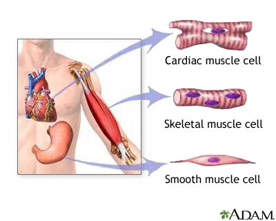
  • Muscle tissue


  • (found at http://www.nlm.nih.gov/medlineplus/ency/images/ency/fullsize/19917.jpg)
    • 3 types
      • Smooth muscle
        • Fibers are spindle-shaped cells
        • Not striated
        • Located in the walls of hollow internal organs
        • Involuntary contraction
      • Cardiac muscle
        • Forms heart wall
        • Striated, tubular, branched
        • Relaxes completely between contractions, preventing fatigue
        • Involuntary contraction
      • Skeletal muscle
        • Supports body
        • Striated
        • Makes bones move; contraction causes movement of bones at a joint
        • Helps maintain constant body temperature
          • Contraction causes ATP to break down
        • Contraction assists movement in cardiovascular and lymphatic vessels
        • Helps protect internal organs and stabilize joints
        • Work in pairs
        • Well organized
          • Contains fascicles (bundles of skeletal muscle fibers)
            • Each fascicle and each fiber of a fascicle are surrounded by connective tissue
        • Covered with connective tissue called fascia, which extends beyond muscle and becomes that muscle’s tendon (connecting to the appropriate nearby bone[s])
  • Muscle fiber cell


(found on slide 6 of Movement – BIO 156 PowerPoint presentation)

    • Large cells, visible with the naked eye
    • Sarcolemma – the plasma membrane that forms T tubules
      • An “excitable” membrane that can generate an action potential, just like neuron membranes do
    • T tubules – extend into muscle fiber, convey impulses causing calcium to be released from the sarcoplasmic reticulum
      • Penetrate into cell, contacts sarcoplasmic reticulum
      • Membrane extensions
      • Crucial in actin-myosin, or muscle, movement
    • Sarcoplasm – muscle cell’s version of cytoplasm; contains organelles, including myofibrils
    • Sarcoplasmic reticulum – made of smooth endoplasmic reticulum (ER) that has expanded portions that store calcium, which is essential for muscle contraction
      • A muscle cell/muscle fiber contracts when calcium leaves the sarcoplasmic reticulum, and relaxed when calcium returns to the sarcoplasmic reticulum.
    • Glycogen – stores energy for muscle contraction; is a polysaccharide
    • Myoglobin – red pigment; stores oxygen for muscle contraction
    • Myofilament – actin filament or myosin filament that are accountable for muscle striations and contractions
      • Thick filament – myosin proteins, shaped like a golf club
      • Think filament – actin proteins; tropomyosin and troponin are important to thin filament because they help myosin bind to actin
      • Sliding filament – movement of actin filament in relation to myosin filament
    • Myofibril – myofilament bundle that contracts
      • Each cell has hundreds of myofibrils
      • Each myofibril contains myofilaments and the actions they perform
  • Motor unit – a nerve fiber and all the muscle fibers it innervates
    • Obeys the all-or-none law
    • A whole muscle usually has many motor units
  • Muscle’s 4 possible energy sources
    • 2 stored in muscles
      • Glycogen
      • Fat (triglycerides)
    • 2 stored in blood
      • Blood glucose
      • Plasma fatty acids
  • Muscle cells – 3 ways they acquire ATP


    • (found on slide 10 of Movement – BIO 156 PowerPoint presentation)
    • Formation of ATP by creatine phosphate (CP) pathway
      • Anaerobic
      • Simplest, most rapid way to produce ATP
      • Consists of one reaction only, makes it very fast transaction
      • Used for high-intensity exercise over 5 seconds or less
    • Formation of ATP by fermentation
      • Anaerobic
      • Produces only 2 ATP
      • Pathway most likely to begin with glycogen
      • Fast-acting
      • Results in lactate buildup
    • Formation of ATP by cellular respiration
      • Aerobic; involves use of oxygen by mitochondria
      • Does not immediately supply ATP for muscle contraction
      • More likely to supply ATP when exercise is submaximal in intensity
  • Muscle twitch – single contraction that last only a fraction of a second
    • 3 stages
      • Latent period
      • Contraction period
      • Relaxation period
  • Muscle fiber – 2 types
    • Fast-twitch muscle fiber
      • Is anaerobic – uses primarily fermentation and CP pathway
      • Has explosive power
      • Fatigues easily
    • Slow-twitch muscle fiber
      • Is aerobic – prefers to use cellular respiration
      • Has steady power
      • Has endurance
  • Calcium – how it is released into muscle
    • 1) “Motor neuron impulse arrives to axonal terminus” (found on slide 7 of Movement – BIO 156 PowerPoint presentation)
    • 2) “Neuro-muscular junction synapse passes message on to muscle cell”; in other words, this is where the message is passed on (quote found on slide 7 of Movement – BIO 156 PowerPoint presentation)
    • 3) Sarcolemma has the action potential passed all along the cell membrane and into the T tubule system
    • 4) This voltage change then causes calcium to be released into the (muscle) cell
    • 5) As a result, the released calcium causes the sarcomeres, or actin-myosin units, to shorten
  • Sliding filament model


    • (found on slide 8 of Movement – BIO 156 PowerPoint presentation)
    • “Rowing” – the myosin, or thick, filaments pull on the actin, or thin, filaments
    • Calcium in sliding filaments
      • Binding calcium allows…
        • Myosin to undergo change (so it will interact with actin)
        • Cycling of molecular cross bridges what changes their shape
    • Uses ATP
    • Here is an image portraying the importance and use of ATP and calcium in sliding filaments


    • (found on slide 9 of Movement – BIO 156 PowerPoint presentation)
    • Muscle cells are lined up in skeletal muscle. When many of them contract simultaneously, the whole muscle shortens and brings about body movement.

Joints

  • “Movement happens across joints between skeletal elements” (from slide 11 of Movement – BIO 156 PowerPoint presentation)
  • “Joints connect skeletal elements” (from slide 13 of Movement – BIO 156 PowerPoint presentation)
  • Joints – 3 types
    • Fibrous – immovable; e.g. sutures between cranial bones
    • Cartilaginous – slightly moveable, connected by hyaline cartilage (e.g. costal cartilage that joins ribs to sternum) or fibrocartilage (e.g. intervertebral disks)
    • Synovial – freely moveable, has a cavity filled with synovial fluid (lubricates joint)


      • (found on slide 13 of Movement – BIO 156 PowerPoint presentation)
      • Generalized synovial joint – e.g. knee
      • Ball-and-socket joins – e.g. shoulder
      • Hinge joint – e.g. elbow
      • “Skeletal elements linked by synovial joints move when muscles pull on those skeletal elements; this is how movement happens” (from slide 13 of Movement – BIO 156 PowerPoint presentation)
  • Technical names for specific movements across joints


  • (found on slide 14 of Movement – BIO 156 PowerPoint presentation)

Bones (and cartilage)

· Skeletal system

o 206 bones in the adult body

o Fibrous connective tissue makes up the ligaments and tendons that hold it together

o 5 functions

§ Supports body

§ Protects soft body parts

§ Produces blood cells

§ Stores minerals and fat

§ Permits flexible body movement when combined with muscle

· Skeleton – 2 groups


· (found at http://www.stpeters.k12.nf.ca/skel.jpg)

o Axial skeleton

§ Skull – cranium, facial boned

§ Hyoid bone – only bone in body that does not articulate with another bone

· Attached to temporal bones and muscle and ligament

· Attached to larynx by a membrane

§ Vertebral column – 33 vertebrae

· 4 curvatures – this makes it stronger than it would be if it were a straight column

§ Rib cage (thoracic cage) – thoracic vertebrae, ribs (12 pairs) and their associated cartilages, sternum

o Appendicular skeleton – bones within the pectoral and pelvic girdles and their attached limbs

§ Pectoral girdle, left and right – scapula and clavicle

· Upper limb

o Arm – humerus

o Forearm – radius and ulna

o Hand – carpals, metacarpals, phalanges

§ Pelvic girdle, left and right – coxal bones

· Lower limb

o Thigh – femur (longest and strongest bone in the body)

o Leg – tibia and fibula

o Foot – tarsals, metatarsals, phalanges

· Bone – a tissue with nerve and blood supplies and cells

o Primary calcium storage site (calcium needed for muscle contraction)

o Compact bone

§ Highly organized

§ Composed into osteons (tubular units)

· Osteocytes live in lacunae

o Tiny chambers in concentric circles around a central canal

· Matrix fills rest of space between

o Spongy bone

§ Unorganized appearance

§ Contains trabeculae (thin plates) that are unequally spaced

§ Spaces filled with red bone marrow

§ Also has osteocytes in trabeculae

· Long bone anatomy


o (found on slide 16 of Movement – BIO 156 PowerPoint presentation)

o Shaft (main part) called diaphysis

§ Large medullary cavity

· Lined with endosteum (thin, vascular membrane)

· Filled with yellow bone marrow (stores fat)

o Expanded region at the end (epiphysis)

§ Composed mostly of spongy bone

§ Coated with thin layer of hyaline cartilage (also called articular cartilage in this case, because it is at a joint)

o Covered with periosteum (fibrous connective tissue)

o Cartilage is at the ends of the bone

o Growth plate – between diaphysis and epiphysis at both bone ends

§ Cartilaginous growth plate – where bone growth occurs


· (found on slide 18 of Movement – BIO 156 PowerPoint presentation)

· 3 cell types associated with bones

o Osteoblasts – bone-forming cells

o Osteocytes – mature bone cells derived from osteoblasts

o Osteoclasts – bone-absorbing cells

· Bone growth

o Ossification – the formation of bone

§ 2 types during skeletal formation in embryonic development

· Intramembranous ossificaction FIND ONLINE PIC

o Bones develop between sheets of fibrous connective tissue

o Develops only flat bones, e.g. skull

· Endochondral ossificiation

o Most human bones formed this way

o Bone replaces cartilaginous bone models


o (found on slide 17 of Movement – BIO 156 PowerPoint presentation)

o 5 steps/parts

§ Cartilage model – made by hyaline cartilage being laid down by chondrocytes in future bone’s shape

· Eventually chondrocytes die off

§ Bone collar – newly formed periosteum makdes osteoblasts

· Osteoblasts secrete organic bone matrix, which begins to calcify

§ Primary ossification center – osteoblasts brough to interior of bone by blood vessels; osteoblasts begin to make spongy bone

· The first bone formation center in the bone

§ Medullary cavity and secondary ossification sites

· Spongy bone of diaphysis is absorbed by osteoclasts

o This results in the medullary cavity

· Secondary ossification centers from in epiphyses shortly after birth

§ Epiphyseal (growth) plate

· Cartilage band between primary ossification center and each secondary ossification center

· 4 layers (starting nearest to the epiphysis)

o Resting zone – where cartilage remains

o Proliferating zone – produces new cartilage cells

o Degenerating zone – cartilage dies off here

o Ossification zone – where bone forms

· Bone remodeling – when bones change in size, shape, and/or strength due to stress; e.g. bone fractures, after which it heals

o 18% of bone is recycled each year in the body

o Keeps bones strong

o Why bones respond to stress: medullary cavity


§ (found on slide 19 of Movement – BIO 156 PowerPoint presentation)

§ Located in diaphysis

§ Hollow tube filled with bone marrow

§ Where blood and nerves are in the bone

§ This structure makes the bone stronger than it would be if it were a solid bone through and through

o Allows body to regulate amount of calcium in the blood

§ Bones store calcium

o 2 hormones regulate blood calcium level

§ Parathyroid hormone (PTH)

· Accelerates bone recycling

§ Calcitonin

· Opposite action of PTH

§ Calcium regulation negative feedback system


o (found on slide 20 of Movement – BIO 156 PowerPoint presentation)

o Osteoporosis – comes from the calcium need outweighing the bone support role

§ Affects elderly, particularly females

§ Happens when bone resorption happens faster than deposition

§ Possibly from low estrogen levels

§ Leads to higher fracture incidence, especially in hip, vertebrae, long bones, pelvis

§ Remedies

· Estrogen treatments tried, and now discouraged because of its link to higher cancer rates

· Keep calcium in the diet, especially green leafy calcium, which is considered better for a person than dairy calcium

o Also, make sure person has enough vitamin D, as it aids body in correctly using its calcium

· Best remedy – lifelong habit of activity (particularly weight-bearing)

§ Here is an image of an osteoporotic bone on the left and a healthy bone on the right


· (found on slide 21 of Movement – BIO 156 PowerPoint presentation)

· Bone repair

o 4 steps

§ Hematoma – after fracture, blood forms clotted blood mass at the space of bone break; takes 6-8 hours

§ Fibrocartilaginous callus – tissue repair beings, fibrocartilaginous callus forms at the break; takes 3 weeks

§ Bony callus – osteoblasts…

· Produce trabeculae of spongy bone

· Convert fibrocartilage callus to bony callus (joins bones together again)

§ Remodeling – osteoblasts build new compact bone at the periphery, while osteoclasts absorb spongy bone

· Cartilage

o Not as strong as bone, more flexible

o Made of chondrocyte cells

§ Are irregularly grouped in lacunae

o Has no nerves or blood vessels

o 3 types

§ Hyaline cartilage – firm, somewhat flexible

§ Fibrocartilage – stronger than hyaline cartilage; able to withstand tension/pressure

§ Elastic cartilage – more flexible than hyaline cartilage

Movement Summary

1. “Neurons trigger muscle to contract based on stimuli or sensory input”

2. “Muscle contracts by sliding filaments”

3. “Muscle pulls on bone to cause movement across joint”


Friday, July 6, 2007

Online Lab Five -- HHMI Virtual Neurophysiology Lab

Katie Meyers

Online Lab Five – HHMI Virtual Neurophysiology Lab

Here is my first screen shot that shows the manipulator with the oscillope trace.

Here is my ultra-violet image of the neuron with dye in it showing it has the shape of a sensory neuron.

1. What is the electrode measuring?

The electrode is measuring the activity of the neuron(s).

2. Why use leeches in neurophysiology experiments?

Their nerve cords have very large and easily accessible neurons on them, making them easier to dissect and experiment on.

3. What is the difference between a sensory and a motor neuron?

A sensory neuron conducts impulses from an organ to the spinal cord and brain. A motor neuron does just the opposite; it conducts impulses from the brain and spinal cord to a muscle or gland.

4. Do you think a leech experiences pain? What is pain?

Pain is the “hurt or suffering of the body or mind” (from http://dictionary.reference.com/browse/pain). As long as leeches have brains of some sort (I am assuming they do, as they are living creatures that act of their own volition), I would say that leeches do feel pain, since this experiment shows that leeches have nerve cords and (large) neurons, which will transmit signals to their brain, thereby making them aware of physical pain.

5. What were the two most interesting things about doing this lab?

I have never dissected before, and I am also easily unsettled at the thought of doing so. However, this lab gave me a relatively realistic dissection experience, without the gore or trying to physically cut something open. Also, as this was my first time dissecting, I was grateful that they did not make it as difficult as it could have been in reality (e.g. they make the incisions without you having to worry whether or not you cut too deep or too shallow). I also really enjoyed figuring out what types of neuron readings I was picking up, and how to tell the types of each neuron I was examining. It was an informative, not very hard, and enjoyable experience.

6. Anything you found confusing or didn't like about the lab?

During the dissection, I did not click the exact place on the screen I was supposed to once or twice. However, I do not see that as the lab’s fault, since the directions were clear. It was just me not being as careful as I should have been. Otherwise, I have no complaints about this lab assignment.

Compendium Review Five -- Nervous Function

Katie Meyers

Compendium Five – Nervous Function

Introduction to the Nervous System

  • Nervous System
    • 2 parts
      • Central Nervous System
        • Spinal cord
        • Brain
      • Peripheral Nervous System
        • Contains nerves
        • Is made up of 2 systems
          • Somatic System
          • Autonomic System
            • Sympathetic Division
            • Parasympathetic Division
        • 3 functions
  • Nervous Tissue – 2 types
    • Neurons
      • 3 types
      • 3 parts
    • Neuroglia

Resting Potential, Action Potential, Synapse

  • Resting potential
  • Action potential
  • Synapse

Sensation

  • Sensation
  • Sensory receptors
    • 2 kinds
    • 4 categories
    • 3 types
  • Specific, or special, senses
    • Taste
    • Smell
    • Vision
    • Hearing
    • Equilibrium

Drug Abuse

  • Drug abuse
  • Drug addiction
  • 6 most popular drugs
    • Alcohol
    • Nicotine
    • Cocaine
    • Methamphetamine
    • Heroin
    • Marijuana

Introduction to the Nervous System

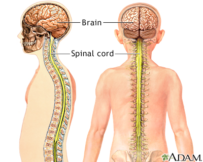
  • Nervous system
    • 2 parts


      • (found at http://www.nlm.nih.gov/medlineplus/ency/images/ency/fullsize/19588.jpg)
        • Where sensory info is received and motor control is initiated
        • Composed of two nervous tissue types – grey matter and white matter
          • Grey matter – contains cell bodies and short, nonmyelinated fibers
          • White matter – contains tracts – bundles of myelinated axons that run together
        • Spinal Cord


        • (found at http://www.infovisual.info/03/img_en/024%20Spinal%20cord.jpg)
          • From brain base through large opening in skull (foramen magnum) and into vertebral canal formed by openings in the vertebrae
          • Provides means of communication between the brain and the peripheral nerves leaving the spinal cord
          • If severed, loss of voluntary control and sensation occurs – called paralysis
          • Center for thousands of reflex arcs
          • Central Canal
            • Contains cerebrospinal fluid
            • Also contains neurons, motor neurons, and interneurons
              • The interneurons communicate with the sensory and motor neurons
          • Grey Matter – shaped like the letter “H”
          • White Matter – occurs around grey matter; contains ascending tracts that take info to the brain, as well as descending tracts that take info from the brain
        • Brain


        • (found at http://www.medem.com/MEDEM/images/ama/ama_brain_stroke_lev20_thebraineffectsstroke_01.gif)
          • Has four ventricles – two lateral ventricles, third ventricle, and fourth ventricle
            • The two lateral ventricles contain the cerebrum
            • The third ventricle contains the diencephalon
            • The fourth ventricle contains the brain stem and cerebellum
          • Cerebrum (also called telencephalon)
            • Largest portion of the human brain
            • Last center to receive sensory input and carry out integration before commanding voluntary motor response
            • Communicates and coordinates with other brain parts
            • Has two halves, or hemispheres (remember that it is located in two ventricles)
            • Sulci (shallow grooves) separate the hemispheres into lobes
              • Four lobes – frontal lobe, parietal lobe, occipital lobe, temporal lobe
              • Each lobe is associated with a particular function
            • Cerebral cortex – gray matter that covers cerebral hemispheres
              • Contains motor, sensory, and associated areas
              • Contains the primary motor and somatosensory areas of the brain
                • Somatosensory area has primary taste, visual, auditory, and olfactory areas
            • Association areas – where integration occurs
              • Visual association area – associates new visual info with previously received visual info
              • Auditory association area – does the same with auditory info as the visual association does with visual info
              • Somatosensory association area – processes and analyzes sensory info from skin and muscles
            • Processing centers
              • Receive info from other association areas
              • Perform higher-level analytical functions
            • Central white matter – makes up much of the rest of the cerebrum
          • Diencephalon
            • Contains hypothalamus and thalamus
              • Hypothalamus – regulates hunger, sleep, thirst, body temperature, and H2O balance; control pituitary gland, therefore serving as the link between the nervous and endocrine systems
              • Thalamus – is two masses of grey matter on the sides and roof of the third ventricle; is the receiving end for sensory input except for smell
            • Also has pineal gland, which secretes melatonin
          • Cerebellum
            • Has two portions, both of which are made up of primarily white matter
            • Receives…
              • Sensory input from eyes, ears, joints, and muscles pertaining to the present position in space of parts of the body
              • Motor output from cerebral cortex, which, after receiving them, sends impulses by the brain stem to skeletal muscles
            • Maintains posture and balance
          • Brain stem
            • Contains midbrain, pons, and medulla oblongata
              • Midbrain – relay system for tracts passing between the cerebrum and spinal cord or cerebellum
              • Pons – literally means “bridge”; contains axon bundles that travel between there cerebellum and the rest of the CNS
              • Medulla oblongata – contains reflex centers for heartbeat, breathing, blood pressure, vomiting, swallowing, coughing, sneezing, and hiccupping
            • Has a reticular formation that is a complex network of grey matter masses and fibers that extend the length of the brain stem
          • Limbic system – a system in the brain that is involved with emotions and higher mental functions
      • Peripheral Nervous System (PNS) – consists of all of body’s nerves


      • (found at http://trc.ucdavis.edu/biosci10v/bis10v/week10/spinalcord.gif)
        • Contains nerves
          • These nerves are composed of axons (the long part of a neuron)
          • All nerves take impulses to and from the CNS
          • Cranial nerves come from the brain, 12 pairs per human
          • Spinal nerves come from the spinal cord and sprout from the sides of the spinal cord, 31 pairs per human
            • Each pair of these spinal nerves serves a specific body area
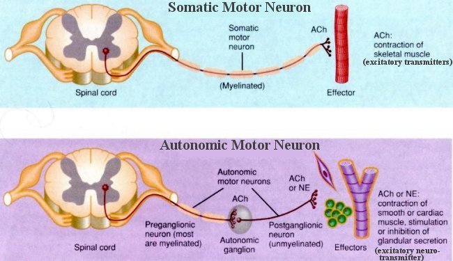
        • Is made up of two systems


        • (found at http://universe-review.ca/I10-13-nerves2.jpg)
          • Somatic system
            • The nerves in this system serve the skin, skeletal muscles, and tendons
            • Take sensory info to and motor commands from these areas
            • Actions are voluntary
            • Reflex arc
              • We do not feel pain until our brain receives and interprets the information about the injury
          • Autonomic system
            • Regulates activity of the cardiac and smooth muscles, as well as the glands
            • Autonomic control – involuntary control, e.g. digestive system, glands, heart, blood vessels; maintains all basic body functions
            • The loss of autonomic function can be fatal
            • Sympathetic and parasympathetic divisions


            • (found at http://www.becomehealthynow.com/images/organs/nervous/sympth_parasymth.gif)
              • Are opposite each other overall
              • Both…
                • Function automatically and usually involuntarily
                • Innervate all the internal organs
                • Utilize two neurons and one ganglion per impulse
              • Sympathetic division
                • “Fight or flight” – accelerates heartbeat, dilates bronchi
                • Inhibits digestive tract
                • Pergangliotic fibers (from the mid-spine) are short
                • Postgangliotic fibers are long when they are in contact with an organ
              • Parasympathetic division
                • “Rest and digest” system – promotes internal responses for a relaxed state
                  • Eye pupils contract
                  • Promotes food digestion
                  • Retards heartbeat
                • Consists of the cranial nerves and fibers from the bottom portion of the spinal cord
                • Pregangliotic fibers long
                • Postgangliotic fibers short
      • These work together and are connected with each other.
    • 3 functions
      • Receives sensory input
      • Processes information (in brain and spinal cord); or, CNS integration – summing up input received from body
      • Motor output – initiating response (muscles result in movement, glands result in secretion); or, CNS generates output as nerve impulses
  • Nervous Tissue – 2 types
    • Neurons – cells that transmit nerve impulses through the body; main cells of the nervous system


    • (found on slide 9 of Nervous Function – BIO 156 PowerPoint presentation)
      • Most neurons never die or divide; once they develop, a human has the same ones for life
      • 3 types
        • Sensory neurons – take nerve impulses from sensory receptors to CNS; detect environmental changes; bring information in
        • Interneurons – all in CNS; receive input from sensory neurons and other CNS interneurons
          • After receiving such input, they sum up the information before communicating with motor neurons
        • Motor neurons – take nerve impulses away from CNS and to effector muscles; send information out
          • Effector muscles – carry out responses to environmental changes, internally and externally
      • 3 parts
        • Cell body – nucleus and other organelles
        • Dendrites – short extension that receives input form sensory receptors or other neurons; connect to other cells, usually neurons or muscle cells
        • Axon – conducts nerve impulses (carries the message); can be very long
          • Long axons have myelin sheath, short ones do not


(found on slide 10 of Nervous Function – BIO 156 PowerPoint presentation)

          • Myelin – “a fat-based insulating substance that isolates neurons and helps speed of action potential propagation” (from slide 10 of Nervous Function – BIO 156 PowerPoint presentation)
            • Schwann cells (a type of neuroglia) – contain myelin in plasma membrane; these cells wrap around axon; only in PNS – CNS uses oligodendrocytes instead
    • Neuroglia – tissue that supports and nourishes neurons

Resting potential, Action potential, Synapse

  • Resting potential – when axon is not conducting an electrical impulse


  • (found on slide 14 of Nervous Function – BIO 156 PowerPoint presentation)
    • Every neuron contains a resting potential
    • -65 to -70 millivolts average (measured by comparison of millivolts inside and millivolts outside cell; -65 means there are 65 less millivolts inside the cell than outside the cell)
      • Sodium-potassium pump prevents diffusion and maintains levels like -65;
      • Pump maintains unequal distribution
        • Greater concentration of sodium ions (Na+) outside axon
        • Greater concentration of potassium ions (K+) inside axon
    • One-third of calories consumed are used to maintain this negative voltage
    • Pumps constantly keep this resting state in preparation for action potential to occur
  • Action potential – rapid messages transmitted by neurons with cell membrane changes; how neurons carry messages


  • (found on slide 15 of Nervous Function – BIO 156 PowerPoint presentation)
    • Ability to sense environment, rapidly transmit messages, and respond
    • “Ability to rapidly carry an ion diffusion mediated change in voltage along the cell membrane” (from slide 13 of Nervous Function – BIO 156 PowerPoint presentation); charged ions cross by diffusion
    • An “all or none” event
    • Always has the same strength, the strength of the reaction is determined by how many nerve impulses are generated in a certain time span
    • Happens only in neurons and muscle cells of mammals
    • Rapid polarity change across the axons of a membrane during the occurrence of a nerve impulse
    • -70 to +40 millivolt comparison in an instant
    • Stimulus causes Na+ and K+ gates to open; the cell’s membrane “depolarizes”, or opens up
      • Na+ opens first, causes +40 millivolt measurement; called depolarization
      • K+ opens last, causes millivolt measurement to return to -65; called repolarization
      • These two steps combined take 3/1000 of a second to occur
    • Refractory period – right after impulse of axon, this ensures sodium gates do not open so action potential cannot move backward
    • Here is an example of an action potential


      • (found on slide 21 of Nervous Function – BIO 156 PowerPoint presentation)
      • Steps in this example
        • 1) “Sensory neurons action potential bring in pain information from skin”
        • 2) “Neurons of spinal cord process information, take decision”
        • 3) “Motor neurons carry output to muscles to move limb away from pain”
        • (from slide 21 of Nervous Function – BIO 156 PowerPoint presentation)
    • As is evidenced by the above example, action potential is a reflex reaction. A person does not even need to think about the situation; the body does it subconsciously, or on its own.
  • Synapse – what happens when action potential arrives at the end of its axon


  • (found at http://www.coolschool.ca/lor/BI12/unit12/U12L04/Synapse.gif)
    • Happens at axon terminal – a small swelling at the end of each axon
    • 3 steps
      • 1) Nerve impulse in axon arrives at axon terminal
      • 2) Calcium enters terminal and stimulates vesicles (in axon terminal that are carrying neurotransmitters) to merge with sending membrane
      • 3) Neurotransmitter molecules released into synaptic cleft, where they then diffuse across the cleft to receiving membrane, where they bind to specific receptor proteins
    • When Na+ is diffused into receiving neuron, it causes excitation.
    • When K+ is diffused into receiving neuron, it causes inhibition.

Sensation

  • Sensation
    • Conscious perception of stimuli
    • Occurs when nerve impulses arrive at the brain’s cerebral cortex
  • Sensory receptors – specialized cells that sense something and interpret what to do, where the signal is coming from, and how “loud” that signal is; dendrites specialized to detect certain stimuli
    • Trigger action potential in connecting sensory neurons
    • Then, signal travels up the spinal cord and to the brain, where the main “Where? What? “How much/how strong?” interpretation and analyzation
    • All over body
    • 2 kinds
      • Exteroceptors – sensory receptors that pick up information from the outside of the body
      • Interoceptors – sensory receptors that pick up information from the inside of the body
    • 4 categories
      • Chemoreceptors – respond to chemical substances in the vicinity, e.g. taste and smell use these
        • Pain receptors (also called nociceptors) – naked dendrites that respond to chemicals released by damaged tissues
      • Photoreceptors – respond to light energy, e.g. eyes
      • Mechanoreceptors – stimulated by mechanical forces, usually resulting in pressure, e.g. hearing, equilibrium, touch
      • Thermoreceptors – stimulated by temperature changes, in hypothalamus and skin
    • 3 types
      • Proprioceptors – mechanoreceptors involved in reflex actions, equilibrium, and posture
        • Help us know position of limbs in space
        • “Proprioception – gives body position by sensing muscle tension” (from slide 27 of Nervous Function – BIO 156 PowerPoint presentation)
          • For example, this allows us to touch our nose with our eyes closed
      • Cutaneous receptors – in dermis; makes skin sensitive to touch, pressure, pain, temperature (all of which are sensations)
      • Pain receptors – in internal organs; sensitive to chemicals released by damaged tissues
    • Sensory brain fields – “For conscious somatic sensory perception, large fields in brain organize information spatially” (from slide 25 of Nervous Function – BIO 156 PowerPoint presentation)


    • (found on slide 25 of Nervous Function – BIO 156 PowerPoint presentation)
  • Specific, or special, senses; only in the human’s head; 5 total
    • Taste


    • (found at http://freda.auyeung.net/5senses/taste.jpg)
      • A limited sensation that uses the tongue
      • About 3,000 taste buds on the human tongue
      • Four main taste sensations – sweet, salty, sour, bitter
        • A possible fifth sensation called umami; considered to taste flavors like “meaty” or “savory”
    • Smell


      • (found on slide 30 of Nervous Function – BIO 156 PowerPoint presentation)
      • Richer than taste; a very fine-tuned sense
      • 80%-90% of taste is due to smell, due to the olfactory cells in the nasal cavity
      • How do we smell? The nose is a very complicated organ that recognizes specific proteins and protein patterns and associates them with specific smells.
    • Vision


      • (found on slide 31 of Nervous Function – BIO 156 PowerPoint presentation)
      • Uses primarily the eyes
      • Another very fine-tuned sense
      • At least one-third of the cerebral cortex takes part in processing visual information
      • How do we see? Light comes into the eyes, which is focused by the cornea. The pupil regulates how much light comes in.
      • Retina
        • “Vision – retina receptors respond to light” (from slide 32 of Nervous Function – BIO 156 PowerPoint presentation)
        • 3 neuron layers
          • Outermost – has rod and cone cells (also known as photoreceptors)
          • Middle – has bipolar cells
          • Innermost – has ganglion cells, whose sensory fibers become the optic nerve
        • Contains the rhodopsin, which changes shape when light hits it
    • Hearing


    • (found on slide 33 of Nervous Function – BIO 156 PowerPoint presentation)
      • Again, a very fine-tuned sense
      • Uses the ears primarily
      • Ear – 3 parts
        • Outer – contains pinna, auditory canal
        • Middle – goes from the eardrum to the bony wall containing the oval window and the round window
        • Inner – contains semicircular canals (helps with equilibrium), vestibule (also helps with equilibrium), cochlea (helps with hearing)
          • Filled with fluid
      • The order in which anatomy is used during hearing – auditory canal, tympanic membrane (eardrum), malleus, incus, stapes, oval window, cochlea, spiral organ, cochlear nerve, brain
    • Equilibrium
      • Responds to body’s movement
      • The anatomy used for equilibrium is in the same apparatus as the inner ear (see above)
      • Achieved and aided by – vesibular nerves, proprioceptors, vision
      • 2 pathways


        • (found on slide 34 of Nervous Function – BIO 156 PowerPoint presentation)
        • Rotational Equilibrium Pathway – uses the three semicircular canals in the ear, which have mechanoreceptors, which detect angular and/or rotational head movement
        • Gravitational Equilibrium Pathway – uses mechanoreceptors in the utricle and saccule that detect vertical and horizontal head movement
          • Utricle and saccule – membranous sacs in the inner ear near the semicircular canals

Drug Abuse

  • Drug abuse – when a drug is taken at does level and under circumstances that increase the potential for harmful effects
  • Drug addiction – when a person tolerates drug and needs more of it for the same effect
    • Can affect fetuses when mother is abusing or addicted to a drug, e.g. a newborn addicted to cocaine
  • 6 most popular drugs that are abused and become addicting
    • Alcohol
      • Is a depressant
      • Harmful effects on body and brain
      • Damages vital body organs, e.g. liver and brain
      • Found in alcoholic drinks (obviously)
    • Nicotine
      • Is a stimulant
      • Found in tobacco, both smoking and chewing
      • Very addictive
      • Damages vital body parts, e.g. lungs
    • Cocaine – “crack”
      • Is a stimulant
      • Derived from the shrub Erythroxylon coca
      • Has euphoric and stimulating effects
    • Methamphetamine – “meth”
      • Is a stimulant – mimics cocaine reactions
      • A synthetic drug made from amphetamine
      • Available in powder or crystal form
      • “Ecstasy”, or methylenedioxymethametaphine, is similar to meth, but without hallucinations
    • Heroin
      • Is a depressant
      • From the resin or sap of the opium poppy
      • Has pain-killing effects
    • Marijuana – “joint”
      • Is a psychoactive, meaning it effects the mind or mental processes
      • Dried parts of the Indian help plant Cannabis sativa

Studying for Bio

Studying for Bio
Me and my dog, Indy