Katie Meyers
Compendium Review Three
Note: If you would like to enlarge a picture, please click on it.
Circulation
- Purpose and performance
- Overview of cardiovascular system
- Blood vessels
- Body’s transportation of objects in/out
- Why cells close to capillaries
- Heart
- Blood pressure
- Lymphatic circulation
Blood, Oxygen, and Cellular Respiration
- Blood cell production
- Blood
- Functions
- Platelets
- Red blood cells
- Plasma
- White blood cells
- Sickle cell
- Cellular respiration
- Oxygen
Immunity
- Three lines of defense
- Two categories of defense
- White blood cells
- Foreign invaders or pathogens
- Clonal selection, or deletion
- Immunity Cells
- Antibodies
- Macrophages
- Acquired immunity
HIV and AIDS
- Origin and discovery
- HIV
- What it does
- Three phases
- What it is made of
- Matrix enzymes
- Replication
- Transmission
- AIDS
- Definition and location
- Opportunistic diseases
- HIV/AIDS geography
Circulation
- Circulation purpose – to help and service the body’s cells
- Performed by the cardiovascular system – heart, blood vessels
- Circulation infiltrates every single bodily structure.
- General overview of the cardiovascular system
- The pump (heart) has big vessels that pump blood into the body.
- The big vessels split up and go into the different main parts of the body – limbs, head, guts, major organs, body wall, skeleton, muscles.
- From here, the medium size blood vessels break up even further into capillaries.
- The oxygen-rich blood expels its oxygen and nutrients out of the capillaries and into the surrounding tissue fluid (or interstitial fluid), which then interacts with the nearby cells and gives them the needed oxygen and nutrients. Right after this is done and the blood is still in the capillaries, the tissue fluid puts cell waste materials (usually CO2) into the blood cells.
- Now the oxygen-deficient blood cells go back to the heart, which then transfers the CO2 waste into the lungs, where it is exhaled.
- After an injury, severed/injured blood vessels heal and reconnect.
- Blood vessels
- Arteries
- Transfer blood away from the heart
- Contains blood that is oxygen-rich, CO2 poor
- Arterial wall’s layers
- Outer layer – connective tissue
- Middle layer – smooth muscle and elastic tissue
- Inner layer – endothelium (thin layer of cells)
- Basement membrane
- Middle artery layer’s muscle contracts, making the artery constrict; if the muscle relaxes, the artery dilates
- The tension of this muscle helps determine blood pressure; e.g., the more dilated the artery is, the lower the blood pressure
- Arterioles – small arteries that are barely visible to the naked eye
- Arterioles branch into capillaries
- Capillaries
- Capillary beds – a network of many capillaries (close together); capillary beds are everywhere in the body
- Beds can be closed or opened by shunts
- Shunts have blood go through the capillary bed or straight from the arteriole to the venule

- Capillaries transfer nutrients and oxygen from the bloodstream into the tissue fluid and nearby cells by diffusion, and vice versa with cellular wastes

- (found on slide 8 of Oxygen – BIO 156 PowerPoint presentation)
- How does blood move through capillaries?
- Two movement controllers
- Hydrostatic pressure – causes H2O to move from capillaries to tissue fluid
- Osmotic pressure – moves substances (waste) from tissue fluid into capillaries
- Created by the presence of salts and plasma proteins

(found at http://training.seer.cancer.gov/module_anatomy/images/illu_capillary_microcirculation.jpg)
- Veins
- Transfer blood to the heart
- Contains blood that is oxygen-poor, CO2-rich
- 70% of blood is in veins at any one time
- Same wall “make” as arteries, except veins have less smooth muscle and connective tissue
- Have valves that open and close to keep blood going only towards the heart
- Skeletal muscle pumps, respiratory pumps, and valves in veins responsible for transporting blood back to heart
- Venules – small veins in which capillary blood goes into; venules join together and become vein
- Picture of overall blood vessel system

(found in slide 7 of Oxygen – BIO 156 PowerPoint presentation)
- “Things” go in and out of body and blood; these following systems are responsible for transporting objects in and out of the body, particularly the blood stream and its circulation system
- Lungs take CO2 out of the body and bring oxygen in
- Intestines/digestive system bring in nutrients
- Kidneys and urinary system take out metabolic wastes; kidneys purify blood of wastes
- Liver picks up amino acids in blood and puts proteins into them; these proteins help transport substances like fats in the bloodstream; the liver also filters out poisons picked up in the intestines
- Here is a basic image of the above systems and their use in circulation

(found on slide 5 of Oxygen – BIO 156 PowerPoint presentation)
o Oxygen – cellular respiration
o Nutrients – cell metabolism
o Immune cells nearby (white blood cells) – eliminate invading microbes
o Waste removal – from cell metabolism
· Heart – circulation’s pump
o Heartbeat is necessary to maintain homeostasis
o Medulla oblongata – brain section that controls internal organs; can alter heartbeat by way of sympathetic and parasympathetic parts of the nervous system; also, hormones epinephrine and norepinephrine control heartbeat rate
o Heart pumps faster when more oxygen is needed
§ Brain, guts, and physically active muscles use a lot of oxygen
· More muscles active, more oxygen needed; explains why heart pumps faster during exercise
o Pulse – how fast heart is pumping
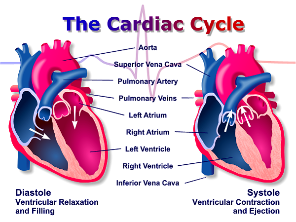
o Cardiac cycle – about 70 heartbeats per minute
§ Two phases
· Systole – contraction phase
· Diastole – relaxation phase
o Majority of heart composed of cardiac muscular tissue – this section is called myocardium
§ Myocardium is serviced by coronary artery and cardiac vein
o Heart surrounded by pericardium – thick sac to support it
o Septum separates heart into left and right sides
o There are four sections in the internal part of the heart – upper left and upper right are called atriums; lower left and lower right are called ventricles
o Heart also has valves to keep blood moving one way in each artery/vein
o Because of valves and four heart sections, oxygen-poor blood and oxygen-rich blood never mix
o Coronary arteries serve the heart by giving it nutrients etc.
o Here is an image of the heart’s anatomy

(found at http://www.anatimation.com/images/heart_diagram.jpg)
o Pulmonary circuit
§ Heart has two pumping systems
· Right side pumps to/from the lungs to collect oxygen and dump CO2
· Left side pumps to/from the body to provide oxygen and nutrients to the body while transporting wastes back from the rest of the body
o Here is an image of the cardiac cycle

(found at http://www.xaraxone.com/FeaturedArt/mar04/assets/images/cardiac_cycle.png)
o The leading cause of death in the Western countries is cardiovascular disease.
· Blood pressure
o Measures the force of blood against vessel walls
o Moves only arterial blood
o Measured by marking two points in the heart cycle
§ Systolic pressure – highest point “as blood is being forced out of heart by contraction of heart muscle” (found on slide 11 of Oxygen – BIO 156 PowerPoint presentation)
§ Diastolic pressure – lowest point, “between heart ‘beats’, when heart is inactive” (found on slide 11 of Oxygen – BIO 156 PowerPoint presentation)
o Sphygmomanometer – measures blood pressure
§ Normal blood pressure measurement is 120/80, systolic pressure/diastolic pressure

(found at http://www.toprol-xl.com/images/sub/inline/normal_vs_high.jpg)
· Lymphatic circulation
o Lymph system – lymphatic vessels collect excess tissue fluid and return it to the cardiovascular system (special blood vessels near the front of the shoulders)
o Lymph – when tissues near tissue fluid collect water, then the water goes into the lymph capillaries and system, therefore becoming lymph fluid
Blood, Oxygen, and Cellular Respiration
· Red bone marrow produces multipotent stem cells, which then can produce red blood cells, white blood cells, plasma, and platelets

(found at http://www.patient.co.uk/showdoc/Pilsinl/183.gif)
· Blood
o Made up of plasma, white blood cells, platelets, and red blood cells

(found at http://www.ndsu.nodak.edu/instruct/tcolvill/435/plasma.gif)
o Blood has three functions
§ Primary transport medium in the body
· Organs and tissues secrete hormones into blood, which transports them to other organs and tissues to influence cell metabolism
§ Defends body against pathogen invasion; blood clots after injury at the site of it
§ Regulatory function
· Regulates body temperature
o Platelets
§ Help blood clot at injuries
§ Result from fragmentation of certain large cells
o Red blood cells
§ Iron, vitamin B12, and vitamin B folic acid are all necessary to produce red blood cells
§ Red blood cell life is about 120 days; they are then destroyed in the liver and spleen
· When a red blood cell is broken down, hemoglobin is released to begin forming new red blood cells
§ Lack a nucleus, have many hemoglobin copies instead
· Hemoglobin – pigment that gives blood its red color
o Contains iron, which accepts oxygen in lungs and releases it in tissue fluid
o One hemoglobin can transport four oxygen molecules
o Directly transports 25% of CO2
o Plasma
§ Made of water, salts, and organic molecules; e.g. plasma proteins
§ Blood cells and cell fragments in plasma
o White blood cells
§ Used for immunity
o Sickle cell
§ Error in DNA code pertaining to hemoglobin formation; a defect in structure of protein molecules – a molecular disease
§ Sickle cell hemoglobin differs from normal hemoglobin by only one amino acid
§ Sickle cells lock together easily due to mutated form; become aggregated
§ Red blood cell sickle cells lose oxygen and warp into sickle shape during high activity; they also become stuck and clot in small blood vessels, causing pain, fever, and swelling; if there is enough tissue damage, death ensues
§ Short term symptoms – out of breath, tires easily
§ Long term symptoms – clogging of capillaries in lungs, kidneys, liver; leads to malfunctioning body systems and death by age 30
§ The only possible cure for sickle cell would be through gene therapy

(found at http://learn.genetics.utah.edu/units/disorders/whataregd/sicklecell/images/sicklecell.jpg)
§ Two forms – sickle cell anemia (severe, two mutant gene copies), sickle cell trait or disease (mild, one mutant gene copy)
· People with sickle cell trait have red blood cells that sickled only when the oxygen/air pressure reached a very low level
§ Higher frequency of sickle cell in people of African descent; this is suspected to be the body’s preventative measure from malaria
· Those with sickle cell (trait or anemia) are immune to malaria
· Those with sickle cell trait do not catch malaria, nor do they develop sickle cell anemia symptoms. As a result, sickle cell trait is more common in Africa, due to evolution.
o Sickle cell trait is defensive against malaria because a person with sickle cell trait’s red blood cells sickle only when infected. As a result, the spleen filters out these sickled red blood cells that happen to contain malaria in them.
· Cellular respiration – usually uses oxygen
o 3 steps
§ Glycolysis – happens with or without oxygen; usually takes a short period of time
§ Citric acid cycle
§ Electron transport chain – transfers across mitochondrion membrane into cell and gives cell the most ATPs
o “Every step of respiration catalyzed by proteins that are coded for in the DNA” (found in slide 14 of Oxygen – BIO 156 PowerPoint presentation)
· Oxygen into blood (occurs in lungs)
o Lung air tubes branch continually until they form small (microscopic) sacs called alveoli, which are surrounded by capillaries
o “Oxygen diffuses across super-thin epithelial tissue of alveolus, across super-thin epithelial tissue of capillary, across red blood cell membrane and is held by hemoglobin protein molecules in red blood cells” (found in slide 15 of Oxygen – BIO 156 PowerPoint presentation)
Immunity
· Immunity is dependent on the cardiovascular system (white blood cells)
· Three lines of defense
o Entrance barriers – skin, mucous membranes
o First responders – e.g. phagocytic white blood cells
o Specific defenses – overcome infection by killing the particular disease
· Two categories of defense
o Nonspecific defenses
§ Barriers to entry
· Skin and mucous membranes
· Chemical barriers – skin oil, perspiration, saliva, tears, urine, acidity of stomach and vagina linings
· Residual bacteria – flora in mouth, intestines, etc.
§ Inflammatory response
· Phagocytic WBCs
o Neutrophils and macrophages – both surround and kill pathogens in body
o Specific defenses – when nonspecific defenses fail; respond to antigens
§ B and T cells – “…recognize the foreign proteins that are on invaders but have been selected so that they don’t recognize (and try to kill) the body’s own cells” (from slide 20 of Oxygen – BIO 156 PowerPoint presentation)
§ Here is an image of specific immune response

(found on slide 26 of Oxygen – BIO 156 PowerPoint presentation)
· White blood cells (WBC)
o Have a nucleus, lack hemoglobin (Hb)
o Fight infections, or invading microbes, as part of immune system
§ Either perform phagocytosis or produce antibodies as immune system defense
o Derived from red bone marrow
o Slightly larger than red blood cells
o Produced more slowly and differently than red blood cells – one WBC can last a human’s lifetime
o Can squeeze through capillary wall into the tissue, and then to the rest of the body
o Two main types of WBC
§ Agranular leukocytes – without granules
· Lymphocytes – recognize invaders, make up 25%-35% of WBCs, specifically immune to pathogens and their toxins
o B cells
§ Produce antibodies and proteins that combine with target pathogens and mark it for destruction
o T cells
§ Cytotoxic T cells – directly destroy pathogen
· Monocytes – largest type of WBC, thought of as cellular “vacuum cleaner” and stimulator of other types of WBC; consume or kill microbes
§ Granular leukocytes – named so because of the granules they contain that carry enzymes and protein that help WBCs defend the body from disease
· Neutrophils – make up 70% of WBCs, have a multi-lobed nucleus, are the first to respond to a bacterial infection, actually consume and kill microbes
· Eosinophils – bi-lobed nucleus, increase in numbers during parasitic infection or allergic reaction
· Basophils – U-shaped lobed nucleus, release histamine and other substances that trigger reactions in other cells

(found at http://www.patient.co.uk/showdoc/Pilsinl/183.gif)
· Foreign invaders or pathogens (bad bacteria and viruses)
o Bacteria
§ Humans are made up of more bacterial cells than human cells
§ Prokaryotic cells
§ Independent cells – metabolically competent
§ Have plasmids, or accessory DNA rings
§ Most life on earth formed by bacterias
§ Most do not cause disease
§ A few do cause disease, and our immune systems recognize such bacteria
§ Some bacteria cells’ walls are surrounded by a capsule
· This allows the bacteria to stick to surfaces
· This also prevents phagocytic WBCs from destroying them
§ Here is an image of the anatomy of a bacterial prokaryotic cell

http://images.encarta.msn.com/xrefmedia/aencmed/targets/illus/ilt/T028556A.gif)
§ Bacteria reproduce by binary fission (see below image)

(found at http://www.brooklyn.cuny.edu/bc/ahp/MBG/MBG3/CB.Binaryfission.GIF)
§ Three different shapes of bacteria
· Bacillus – rod
· Spirillum – curved
· Coccus – spherical

(found at http://www.cfsan.fda.gov/~dms/a2z-b3.gif)
§ A few well-known bacterial sicknesses – strep, food poisoning, gangrene, syphilis
o Viruses
§ “…are escaped parts of genome or DNA of different organisms. They cannot live independent of the cells they escape from.” (from slide 19 of Oxygen – BIO 156 PowerPoint presentation)
§ Not composed of cells; rather, they are obligate parasites
§ Four times smaller than a bacterium
§ Cause sicknesses like – colds, measles, polio, AIDS
§ Made of two parts
· Outer capsid of protein units
· Inner core of nucleic acid
§ Here is an image of two different types of viruses

(found on slide 19 of Oxygen – BIO 156 PowerPoint presentation)
§ Some viruses continually change shape, which makes them harder to recognize, treat, and get rid of.
§ Reproduction
· Can reproduce with just their RNA or with DNA
· Relies on host cell for reproduction

(found at http://cache.eb.com/eb/image?id=95417&rendTypeId=34)
· Clonal selection, or deletion
o “During fetal/childhood/adolescent development, immune cells are exposed to body’s own cells and the proteins that they have on their surface. This happens mostly in the thymus and bone marrow (both of which regulate immune development). Those immune cell precursors that attack our own cells are eliminated. Those that recognize other cells continue to develop as T-cells and B-cells.” (found on slide 20 of Oxygen – BIO 156 PowerPoint presentation)
§ B and T cells recognize antigens with antigen receptors, which are the proteins in the plasma membrane.
· Immunity cells
o B cells
§ Two types
· Plasma B cells – produce specific antibodies
· Memory B cells – ready to produce future antibodies

(found on slide 21 of Oxygen – BIO 156 PowerPoint presentation)
§ Characteristics
· Antibody-mediated immunity against pathogens
· Recognizes presence of specific antigens (foreign bodies) and then undergoes clonal selection
· Clonal expansion produces antibody-secreting plasma cells and memory B cells
· Production, clonal selection, and maturing of B cells all occur in red bone marrow during the first years of life
· B cells have B cell receptors (antibodies) that pick up on one type of antigen
o Then, B cell with its antibody copies to make B cell clones to fight the certain antigen the antibody recognizes
§ Many varieties of B cells are produced by random recombination of genes for different body regions
§ Here is an image of B cell actions

(found on slide 27 of Oxygen – BIO 156 PowerPoint presentation)
o T cells
§ Regulate immune response
§ Three types
· Cytotoxic T cells – kill virus-infected cells and cancer cells – responsible for cell-mediated immunity
o Autoimmune disease – when cytotoxic T cells attack self; no cure yet
· Helper T cells – regulate immunity, control immune response by releasing cytokines to stimulate other immune system cells – do not fight directly
o Cytokines – signaling molecules in T cells, macrophages, and elsewhere
§ Regulate WBC functions
§ Possible use for treating cancer and AIDS
· Memory T cells – ready for future kills
§ T cells have TCRs (T Cell Receptors) in the same way that B cells have BCRs
§ T cells need help recognizing antigens – antigen-presenting cells like macrophages help them do this
§ T cells recognize the difference between “self” and “foreign” antigens – know which ones to destroy (“foreign”)
§ Characteristics
· T cells are cell-mediated immunity against virus-infected and cancer cells
· Produced in bone marrow, mature in thymus
· Antigens must be presented in the groove of an HLA (human leukocyte antigen) molecule
§ Here is an image of T cell actions

(found on slide 27 of Oxygen – BIO 156 PowerPoint presentation)
· Antibodies

(found at http://www.biology.arizona.edu/IMMUNOLOGY/tutorials/antibody/graphics/antibody.gif)

(found at http://www.nwfsc.noaa.gov/hab/habs_toxins/marine_biotoxins/detection/images/AntibodyMolecule.jpg)
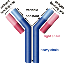
o Very variable proteins that B cells produce in order to recognize antigens (foreign proteins on invaders)
o Proteins made by transcription and translation of certain parts of DNA
o Can be either free in the blood stream or on B cells (antibodies are called BCRs when they are on B cells)
§ T cells also make variable proteins to recognize antigens called TCRs
o 5 classes – IgG (main one in blood), IgM, IgA, IgD, IgE
o Question: “How can each cell have its own different antibody if they all have the same DNA?” (from slide 31 of Oxygen – BIO 156 PowerPoint presentation)
§ Answer: “Recombining regions of DNA that make the antibody recognition site produces all the billions of different combinations of antibodies (and BCRs, TCRs) that recognize any possible invader by the proteins it has on its surface (hopefully).” (from slide 31 of Oxygen – BIO 156 PowerPoint presentation) In other words, DNA contains enough information to make all the different types (millions) of BCRs needed.
· This answer was discovered by Susumu Tonegawa, who won the Nobel prize for his discovery.

(found on slide 31 of Oxygen – BIO 156 PowerPoint presentation)
· Macrophages
o Different types
§ Langerhans – cells in skin
§ Phagocytes – cells in blood
§ Microglial – cells in central nervous system
o Phagocytes help get rid of invading microbes – the following is an example of non-specific immunity or defense
§ First, they move through blood and lymph, then into connective tissues

(found on slide 24 of Oxygen – BIO 156 PowerPoint presentation)
§ Then, they engulf and dissolve the microbes

(found on slide 25 of Oxygen – BIO 156 PowerPoint presentation)
· Acquired immunity – artificial immunity
o Immunization
o Prepared antibodies/immune cells given to a person to combat a disease; this is a temporary treatment
HIV and AIDS
· Originated in Africa, spread to U.S.A. and Europe in the 1950s by the Caribbean
· HIV connection to AIDS was discovered in the 1980s
· AIDS name invented in 1982
· HIV – Human Immunodeficiency Virus – an infection
o The virus that causes AIDS
o Two forms – HIV-1 and HIV-2
§ HIV-1 more widespread and virulent; stimulates cytotoxic T cells
§ HIV-2 stimulates B cells
o HIV infects and destroys immune system cells like helper T cells and macrophages – when these decline, the immune system’s effectiveness declines too
§ Helper T cells are infected, so they send off a warning signal that an infection has entered the body. However, other immunity cells destroy helper T cells because they are infected. As a result, all helper T cells are eventually killed, omitting a crucial part of immune response (the “alarm system”). In the end, it is an opportunistic sickness that takes hold of the body and kills the infected person because their body cannot ward off sicknesses without their helper T cells.
o Three phases of HIV infection
§ Acute – no symptoms, highly contagious, CD4 cell count equal to or greater than 800 cells/mm3
§ Chronic – 200-499 cells/mm3, impaired immune system symptoms
§ AIDS – less than 200 cells/mm3 or development of one or more opportunistic infection/disease
o Originated in Africa, when the natives ate primates that were carrying HIV-1 and HIV-2
o Made of two single RNA strands, various proteins, and an envelope acquired from the host cell

(found at http://www.mcld.co.uk/hiv/images/hivStructure.gif)
o Three types of enzymes are in the HIV’s matrix, which helps protect its genetic material
1. Reverse transcriptase – “catalyzes reverse transcription, which is the conversion of the viral RNA to viral DNA”
2. Integrase – “catalyzes the integration of viral DNA into the DNA of the host cell”
3. Protease – “catalyzes the breakdown of the newly synthesized viral polypeptides into functional viral proteins”
· The three above definitions were quoted from “Human Biology”, by Sylvia S. Mader, 10th ed. page 348
o Must get into a host cell to replicate
o Once in a host cell, HIV is part of the host cell’s RNA (by reverse transcription) and can be copied hundreds of times until the cell bursts from so many copies
o Here is a more detailed, step-by-step explanation of the HIV life cycle
§ 1) Attachment – HIV binds to host cell
§ 2) Fusion – HIV fuses with plasma membrane, virus enters cell
§ 3) Entry – RNA and viral proteins released into the cell
§ 4) Reverse Transcription – reverse transcriptase converts HIV RNA into DNA
§ 5) Integration – viral DNA migrates to nucleus, then viral DNA spliced into cell’s DNA – HIV now provirus: part of cell’s genetic material
§ 6) Biosynthesis and Cleavage – when provirus is activated by different infections, cell produces viral RNA which is transcribed into protein synthetic polypeptides; protease cuts up the long polypeptide chain into smaller pieces
§ 7) Assembly – capsid protein, viral enzymes, and RNA assembled into new viral particles
§ 8) Budding – virus acquires envelope (made of host cell’s plasma membrane) and envelope marker

(found on slide 31 of Oxygen – BIO 156 PowerPoint presentation)
o HIV transmission
§ HIV is a virion (loose virus) that does not live long outside of body fluid, so it is most commonly transmitted through body fluid contact by sexual contact, needle sharing, and blood transfusion
§ HIV not transferred through casual contact
§ HIV most fatal STD (sexually transmitted disease)

(found on slide 32 of Oxygen – BIO 156 PowerPoint presentation)
· AIDS – Acquired Immune Deficiency Syndrome – a syndrome
o Found in many homosexual men in the U.S.A.
o Is the advanced stage of an HIV infection
o The real AIDS epidemic is in sub-Saharan Africa, where it is spreading uncontrollably
o AIDS manifests fully usually when an opportunistic disease takes hold of the patient
o Common opportunistic diseases that cause AIDS deaths
§ Pneumocystis jiroveci pneumonia (PCP) – leading cause of AIDS deaths
§ Myobacterium tuberculosis
§ Toxoplasmic encephalitis
§ Kaposi’s sarcoma
§ Invasive cervical cancer

(found at http://www.cas.muohio.edu/~stevenjr/images/aids98_conditions.gif)
o Is an AIDS vaccine possible? Hopefully one day, but as it is very difficult to find a vaccine that works due to HIV’s high mutation rate (making it hard to keep up with), it will be a long and difficult road to find a real cure, not just medications.
· HIV/AIDS geography
o 75% in Africa; #1 affected region, especially in sub-Saharan Africa
o India has highest number of people with HIV
o Cases are down in numbers in the U.S.A.; must not get complacent in protection efforts
o Over 1% human adults infected with HIV

(found at http://www.agen.ufl.edu/~chyn/age4660/lect/lect_16/aids_countries.jpg)