Thursday, July 19, 2007

Compendium Review Seven -- Reproduction

Katie Meyers

Compendium Review Seven – Reproduction

Human Life Cycle

  • Overview of events in human life cycle
  • Aging

Reproductive Organs (Genitals)

  • 5 functions
  • Male reproductive system
    • Overview of male reproductive organs
    • Steps of male reproductive system
    • Ejaculation
    • Testes
  • Female reproductive system
    • Organs
    • External genitals
    • Meiosis
    • Female hormones
    • Uterine cycle – non-pregnant
    • Ovarian cycle – non-pregnant
    • Uterine and ovarian cycles – pregnant
    • Menopause

Birth Control Options, Infertility, and STDs

  • Birth Control – some examples
  • Infertility
  • STDs – sexually transmitted diseases
    • Viral
    • Bacterial
    • Prevention

Fetal Development at Birth

  • Fertilization
  • Implantation
  • Processes of Development
  • Extraembryonic membranes
  • Development stages
    • Pre-embryonic development
    • Embryonic development
    • Fetal development
  • Genital development
  • Pregnancy and birth
    • Parturition
    • Placenta
    • Mother/fetus blood never mixes
    • Umbilical cord
    • Maternal changes in female anatomy
    • How mother can prevent birth defects
    • Birth
      • 3 stages
    • Many different approaches

Human Life Cycle

  • Overview of events in human life cycle (in order)
    • Meiosis
      • “Reduction division”
      • Occurs only in sex organs
    • Fertilization
    • Fetal Development
    • Birth
    • Childhood/Adolescence
      • Puberty – when a child becomes, by a series of event, a sexually competent adult
        • Reproductive system does not fully function until puberty is complete
        • Occurs…
          • 11 – 13 in females
          • 14 – 16 in males
    • Adulthood


    • (found on slide 4 of Reproduction – BIO 156 PowerPoint presentation)
      • During this stage, adults’ sex organs perform meiosis in preparation of having children
    • Aging
    • Death
  • Changes to each of these stages is due to the aging process

Reproductive Organs (Genitals)

  • 5 functions
    • 1)
      • Males – produce sperm within testes
      • Females – produce eggs within ovaries
    • 2)
      • Males – nurture, transport, and eject sperm
      • Females – transport eggs to uterus
    • 3)
      • Males – penis delivers sperm to…
      • Females – …vagina – takes in sperm, ejects menstrual fluid, is birth canal
    • 4)
      • Females – uterus allows fertilized egg to develop and be nourished within a female
      • Females – breasts provide nourishment after birth
    • 5)
      • Both – testes/ovaries produce respective sex hormones
        • These hormones bring about masculinization/feminization
        • Females – allow pregnancy to continue
  • During sexual intercourse, both male and female will ideally experience orgasms
  • Male Reproductive System
    • (Overview of) Male Reproductive Organs


      • (found on slide 6 of Reproduction – BIO 156 PowerPoint presentation)
      • Testes – produce sperm and sex hormones; primary sex organ; plural (there are two)
        • Millions of sperm in just a few drops of testicular fluid
      • Scrotum – sacs that suspend testes (two of them)
      • Epididymides – ducts; store sperm; this is where sperm also matures
      • Vasa deferentia – conduct and store sperm
      • Seminal vesicles – contribute nutrients and fluid to semen
      • Prostate gland – contributes fluid to semen
      • Urethra – conducts sperm
      • Bulbourethral glands – contribute fluid containing mucous to semen
      • Penis – sexual intercourse organ
      • Prepuce – part of penis; removed during circumcision
    • Steps of the male reproductive system
      • Sperm is produced in left and right testes
      • The vas deferens (left, right) carry it up into the abdominal cavity
      • At base of penis, it (sperm) joins with urethra
      • Semen produced by prostate and seminal glands at the penis’s base
      • Ejaculation occurs
        • When sperm travel from testes, are joined by semen, and ejected through erect penis
    • At time of ejaculation, sperm leaves penis by means of semen
      • 3 types of vesicles/glands add secretions to seminal fluid (see above: seminal vesicles, prostate gland, bulbourethral glands)
      • Seminal fluid
        • Each component is different and has a particular function
          • Basic solution (more viable)
          • Sugar fructose (provide sperm with energy)
          • Prostaglandins (chemicals that contract uterus, moving sperm toward egg)
    • Testes
      • Begin developing inside abdominal cavity, descend into scrotal sacs during last two months of fetal development
      • Scrotum regulates their temperature by holding them closer/farther from body, depending on their current temperature and the environment
      • Each composed of…


        • (found on slide 7 of Reproduction – BIO 156 PowerPoint presentation)
        • Lobules
          • Contain one to three seminiferous tubules
            • Packed with cells undergoing spermatogenesis (production of sperm) by meiosis
              • Seroti cells support, nourish, and regulate spermatogenesis
              • Sperm not ejaculated is reabsorbed by testicular tissue
              • It takes approximately 74 days for sperm to develop from spermatogenesis into sperm
                • Mature sperm (spermatozoa) – 3 parts
                  • Head – nucleus covered by acrosome (which stores enzymes to penetrate egg)
                  • Middle piece – has mitochondria that provide energy for tail movement
                  • Tail – flagellum
                  • Do not live more than 48 hours in female genital tract
        • Interstitial cells
          • Live between seminiferous tubules
          • Secrete sex hormone
            • Most important sex hormone is testosterone


    • (found on slide 9 of Reproduction – BIO 156 PowerPoint presentation)
      • Ovaries – produce eggs (oocytes) and sex hormones (estrogen and progesterone)
        • Site of initial meiosis, where unfertilized eggs are produced
      • Oviducts – conduct eggs; location of fertilization in uterine/fallopian tubes specifically; lined with cilia
      • Uterus (womb) – houses developing fetus
        • Placenta – sustains development of embryo/fetus
      • Cervix – contains opening to uterus
        • Hysterectomy – removal of cervix; a form of sterilization
      • Vagina – receives penis during sexual intercourse
        • Birth canal
        • Exit for menstrual flow
        • Acidic environment
    • External genitals
      • Collectively known as the vulva
        • 2 labia majora (folds of skin)
        • Mons pubis
        • Labia minora – similar to labia majora, just closer to vagina
          • Urethra and vagina openings inside labia minora
        • Glans clitoris
        • Hymen – partially closes vagina; ring of tissue (virginity)
    • Meiosis
      • Occurs in ovaries
      • Phase One
        • Occurs in fetal ovary
        • One egg per month (ovulation part of menstrual cycle) matures and bursts from ovarian wall and taken up into the fallopian tube
      • Phase Two
        • Occurs moment egg is fertilized, if that even happens
      • Possibly very large gap between phases
    • Female hormones
      • Ovaries’ production of hormones


        • (found on slide 10 of Reproduction – BIO 156 PowerPoint presentation)
        • Primary follicles – produce estrogen
        • Secondary follicles – produce estrogen and some progesterone
        • Corpus luteum – produces progesterone
      • Hypothalamus – same function in females as in males
        • Hormone secretion (GnRH, FSH, LH) not constantly present, but secreted at different rates during menstrual cycle
      • Estrogen – responsible for secondary female sex characteristics, e.g. body hair, fat distribution
      • Estrogen and progesterone both required for breast development
    • Uterine cycle – non-pregnant – average 28-day cycle
      • Menstruationdays 1-5
        • Endometrium breaks down
      • Proliferative phase – days 6-13
        • Endometrium rebuilds
      • Secretory phase – days 15-28
        • Endometrium thickens
        • Glands become secretory
    • Ovarian cycle – non-pregnant – average 28-day cycle (occurs simultaneously with uterine cycle)
      • Follicular phase – days 1-13
        • Follicle maturation occurs
        • Estrogen secretion prominent
      • Ovulation – day 14
        • LH spike occurs
      • Luteal phase – days 15-28
        • LH secretion occurs
        • Corpus luteum forms
        • Progesterone secretion prominent
    • Here is a picture of both the uterine and ovarian non-pregnant cycles


    • (found on slide 11 of Reproduction – BIO 156 PowerPoint presentation)
    • Here is a picture of both the uterine and ovarian pregnant cycles – unlike non-pregnant cycle, progesterone does not drop (because it helps maintain uterus wall in preparation of pregnancy)


    • (found on slide 12 of Reproduction – BIO 156 PowerPoint presentation)
    • Menopause – when ovarian cycle stops
      • Begins between the ages of 45 and 55

Birth Control Options, Infertility, and STDs

  • Birth Control – some examples


    • (found on slide 13 of Reproduction – BIO 156 PowerPoint presentation)
    • Abstinence – most reliable
      • Prevents transmission of STDs
    • Family planning – on of the least effective options
      • 70% family planners succeed with the program
    • Specifically female
      • Contraceptives – medications, e.g. birth control pills
      • Contraceptive implants – capsule that utilizes synthetic progesterone, prevents ovulation
      • Contraceptive injections – either progesterone only or a progesterone/estrogen mix
        • From a few weeks to three months between shots
      • Contraceptive vaccines – immunize woman to hormones associated with egg production, e.g. HCG
      • “Morning-after pills” – emergency contraception
      • Intrauterine device (IVD) – small molded plastic piece inserted into uterus by physician
        • Mostly inhibits fertilization
        • Makes implanting impossible
      • Diaphragm – soft latex cup with flexible rim
        • Lodges behind pubic bone, fits over cervix
        • Individually fitted
        • Used with spermicidal jelly or cream
        • Put in no more than two hours before sexual activity
        • Remove at least six hours after sexual activity
      • Female condom – large polyurethane tube
        • Open end has ring that covers external genitals
        • Considered a barrier method
          • Renewed interest in STD protection possibility
      • Tubal ligation – cuts and seals oviducts
        • Brings about sterilization
        • Should be considered permanent
    • Specifically male
      • Male condom – latex sheath goes over erect penis
        • Better form of protection if used with spermicide
        • Same as female condom in that it is considered a barrier method
          • Renewed interest in STD protection possibility
      • Vasectomy – cuts and seals vas deferens


        • (found on slide 8 of Reproduction – BIO 156 PowerPoint presentation)
        • Form of sterilization
        • Should be considered permanent
  • Infertility
    • Definition – failure of a couple achieving pregnancy after one year of effort (unprotected, regular intercourse)
    • Estimated 15% of couples infertile
      • 40% -- males; low sperm count or much abnormal sperm
      • 40% -- females; body weight
      • 20% -- both
    • Assisted reproductive technologies
      • Artificial Insemination by Donor – sperm placed into vagina by physician
      • In Vitro Fertilization – conception in lab glassware
      • Gamete Intrafallopian Transfer – similar to IVF
        • Eggs removed and reintroduced in same procedure/time period
      • Surrogate mothers – paid women bring stranger’s baby to term
      • Intracytoplasmic sperm injection
        • Used in sever male fertility problems
        • One sperm directly injected into egg
  • STDs – sexually transmitted diseases
    • Viral – none curable yet
      • HIV infection – fatal
      • Genital warts
        • Caused by human papillomavirus (HPV)
      • Genital herpes
        • Caused by herpes simplex virus
          • Type One – cold sores, fever blisters
          • Type Two – genital herpes
      • Above two very common and uncomfortable
      • Hepatitis
        • Infects liver by failure, cancer, or death
        • Easily transmittable
        • No immediate consequences
        • ABCDEG
          • A – not sexually transmitted; fecal to mouth
          • BCDEG – transmitted by…
            • Sexual contact
            • Contaminated blood
          • B – most common sexually transmitted hepatitis; can lead to liver failure
          • CDEG – liver cancer possibilities; no vaccines yet available
    • Bacterial – curable with antibiotics
      • Chlamydia
      • Gonorrhea – 40% strains now resistant to antibiotic therapy
      • Syphillis – 3 stages
      • Bacterial vaginosis (BV)
        • Responsible for about 50% of vaginitis in American women
        • “Caused by a disruption of the normal flora in the vagina leading to an overgrowth of certain bacteria” (from slide 16 of Reproduction – BIO 156 PowerPoint presentation)
      • Candida albicans
        • Overgrowth of normal yeast in vagina
        • Characterized by – tissue that is red, inflamed, itchy; with, curdy discharge sometimes too
        • Women using birth control hormones/antibiotics more prone
      • Trichomonas vaginalis
        • Caused by a type of protozoan
        • Causes – frothy discharge, foul smell, itching
        • Common cause of vaginitis
    • Ways to prevent STD transmission
      • Abstinence
      • Long-term monogamous relationship(s)
      • Be aware of partner’s STD history and risky (STD-wise) behavior
      • Practice safe sex

Fetal Development and Birth

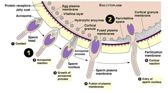
  • Fertilization – union of sperm and egg resulting in zygote


  • (found at http://www.bio.davidson.edu/Courses/Molbio/MolStudents/spring2005/Dresser/sperm%20and%20egg%20fusion.jpg)
    • Flagellum on sperm finds (swims towards) egg
    • Sperm head only (nucleus) fuses with egg’s nucleus
    • Zygote receives cytoplasm and organelles from mother only
    • Approximately one week from fertilization to implantation
      • During that week, only mitosis occurs
  • Implantation
    • Normally in uterine wall
    • Ectopic pregnancy – when a fertilized egg implants before getting to uterine wall, e.g. oviduct
      • Can be dangerous, lead to hemorrhaging
    • First step to take if pregnancy is suspected – find where zygote implanted
  • Processes of Development
    • Cleavage – occurs during mitotic cell division of the zygote
    • Growth – cell division accompanied by daughter cells becoming larger
    • Morphogenesis – shaping of the embryo
      • First evident when some cells move/migrate compared to the other cells – such movements make embryo assume various shapes
    • Differentiation – when cells take on specific structure and/or function
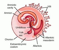
  • Extraembryonic membranes


  • (found at http://img.tfd.com/dorland/thumbs/amnion.jpg)
    • Outside embryo
    • 4 types
      • Chorion – develops into fetal half of placenta
      • Allantois – extends away from embryo
        • Deal with kidneys/urinary tract and has umbilical blood vessels
      • Yolk sac – first to appear of the four
        • Contains many blood vessels – first site of blood cell formation
      • Amnion – enlarges relative to embryo/fetus
        • Contains cushioning fluid to protect embryo/fetus
  • Overview of early embryonic development (about first month)
    • “Basic body plan laid down”
    • “No organs formed”
    • “Placenta develops”
    • “Very little growth, fetus still tiny”
    • Above quotes from slide 20 of Reproduction – BIO 156 PowerPoint presentation
  • Development Stages
    • Pre-embryonic development
      • Contains the events of the first week
        • Zygote division while traveling down oviduct to uterus


    • (found on slide 19 of Reproduction – BIO 156 PowerPoint presentation)
    • Embryonic development


      • (from slide 20 of Reproduction – BIO 156 PowerPoint presentation)
      • Goes from second week to end of second month
      • Week two – embryo begins implanting in uterus
        • Gastrulation occurs
          • Turns inner cell mass into embryonic disk
          • An example of morphogenesis
          • At completion, there is…
            • 3 primary germ layers


              • (found on slide 21 of Reproduction – BIO 156 PowerPoint presentation)
              • Ectoderm
              • Mesoderm
              • Endoderm
            • Embryonic disk has become embryo
      • Week three – two important organ systems appear
        • Nervous system
        • Heart (both third and fourth weeks)
      • Week four – body stalk (future umbilical cord) connect embryo to chorion; limb buds (flippers) appear
      • Week five – head enlarges; sense organs gain prominence; visual development (possible to see) of eyes, ears, and nose
      • Week six to week eight – embryo changes into recognizable human being; head achieves normal relationship with body; neck region develops; nervous system responsive (to reflex actions); all organ systems established
    • Fetal development – fetus is recognizably human
      • Month three – fingernails appear; gender distinguishable by ultrasound
      • Month four – skeleton visible; hair begins appearing; cartilage begins being replaced by bone
      • Month five – heartbeat heard; vernix caseosa (cheesy protective coating) begins being deposited; mother begins feeling movment – pregnancy obvious
      • Month six – body covered with lanugo (fine hair); wrinked and reddish skin
      • Month seven – testes descend into scrotum; eyes are open; possible for baby to survive if born now
      • Month eight – body hair begins disappearing; depositing of subcutaneous fat begins
      • Month nine – fetus ready for birth and about 20 ½ inches long and 7 ½ pounds
    • Male and female development of genitals


    • (found at http://www.mie.utoronto.ca/labs/lcdlab/biopic/fig/41.13.jpg)
      • Development of gonads and ducts, development of external genitals
      • Sex of fetus determined at fertilization
        • Males – XY
          • Male XX syndrome
        • Females – XX
          • Female XY syndrome
        • Both syndromes result in ambiguous sex determination
        • Gonads do not start developing until the seventh week
        • At six weeks, males and females have same types of ducts
        • At fourteen weeks, primitives testes/ovaries are located deep inside abdominal cavity
  • Pregnancy and birth
    • Parturition – process of birthing offspring
    • Placenta – source of progesterone and estrogen during pregnancy
      • 2 sides
        • Fetal side – contributed by chorion
        • Maternal side – consisting of uterine tissue
      • Placental hormones cause…
        • Fluctuations in energy levels
        • Uterus relaxation
        • Pulmonary values to increase
        • Stress incontinence
        • Decreased venous return
        • Edema
        • Varicose veins
        • Stretch marks (Striae gravidarum)
        • Peptide hormone (from placenta) brings about pregnancy-induced diabetes
    • Mother/fetus blood never mixes


    • (found at http://images.main.uab.edu/healthsys/ei_0181.gif)
    • Umbilical cord – fetus’s lifeline
      • Contains umbilical arteries and vein
    • Maternal changes in female anatomy
      • Shift in organs
        • Bladder and rectum pushed superiorly and squeezed, resulting in changes in urination and defecation patterns
        • Stomach is compressed, making the mother eat more often, but less at every meal
        • Appetite change due to immune response changing to avoid possible poisons for the fetus’s sake
      • “Postural changes to compensate for anterior weight” (found on slide 22 of Reproduction – BIO 156 PowerPoint presentation)
      • Breasts enlarge because of milk production
        • First milk production begins around the sixth month of pregnancy
    • How mother can prevent birth defects
      • Get a physical exam
      • Have good health habits
      • Avoid…
        • Drinking alcohol
        • Smoking cigarettes
        • Taking illegal drugs
        • Taking medications not approved by your physician
        • Exposure to environmental toxins and radiations
      • Help prevent conditions associated with birth
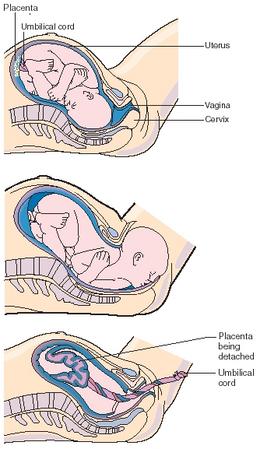
    • Birth
      • 3 stages

      • (found at http://www.scienceclarified.com/images/uesc_02_img0089.jpg)
        • Stage One
          • Effacement – taking up the cervix – done by uterine contractions
          • Amniotic membrane ruptures, if it has not already
          • First stage of parturition ends once cervix is completely dilated
        • Stage Two
          • Beings when uterine contractions occur every one to two minutes and last about one minute each
          • Mother has desire to push, or bear down
          • As soon as head is delivered physician may hold head and guide it downward
          • After birth, umbilical cord is cut and tied once baby is breathing normally and functioning independently
        • Stage Three
          • Afterbirth (placenta) is delivered about 15 minutes after delivery
    • Many different approaches to the birthing and the child-raising processes

Wednesday, July 18, 2007

Online Lab Seven -- Fetal Development: 10 Stages

Katie Meyers


Online Lab 7: Fetal Development – 10 Stages


NOTE: The format of these descriptions will be as follows:
  • stage “title”
    • quick description
    • when it occurs
    • why I think it is a significant stage
  • Neural folds and heart folds begin to fuse.
    • Neural folds coming from neural tube begin to fuse; heart tube takes on S-shape, which establishes asymmetry of heart
    • 21-23 days post-ovulation
    • Cardiac muscle contraction begins; eye and ear cells are present; simplified (early) nervous system begins developing

    • Two Pharyngeal Arches Appear
      • S-shaped embryo, bulb-like tail, connective stalk to placenta; primitive S-shaped heart is beating, peristalsis begins; primary blood vessels along central nervous system are connecting
      • 23-25 days post-ovulation
      • Heart beating, peristalsis present
      • Future Cerebral Hemispheres Distinct
        • Brain enlarges by one-third; external ears begin developing; separate nasal pits; closed lens pit; four pairs of pharyngeal arches visible; distinct regions of upper limb bud distinct; innervation begins in lower limb buds
        • 35-38 days post-ovulation
        • Cerebral hemispheres are distinct
      • Four-Chambered Heart
        • Jaw/facial muscles develop; olfactory (sense of smell) bulb develops in brain; teeth buds form; pituitary begins forming; heart separates into four separate chambers; diaphragm forms; germ cells determining gender arrive at pelvis; digist more distinct
        • 42-44 days post-ovulation
        • Heart divides into four chambers; smell develops; pituitary begins forming; gender germ cells arrive at pelvis

      • First Detectable Brain Waves
        • Brain has first detectable brain waves; inner ear canals start forming; gonads form; toenails begin to appear; joint more distinct; critical period for lower limbs is almost over; muscle develops
        • 48-51 days post-ovulation
        • First detectable brain waves
    • Cartilage Begins to Transform Into Bone
      • Head develops fissure human characteristics; brain can move muscles; end of critical heart development period; clitoris/penis begins to form; hands and feet approach each other; primary ossification centers appear in long bones (directing replacement of cartilage with bone)
      • 54-56 days
      • Cartilage begins to transform into bone (beginning with primary ossification centers)
    • Fetus Begins to Move Around
      • Head rests on neck, not shoulders; sucking mouth muscles fill cheeks; salivary glands begin functioning; discernible hair pattern; heartbeat detectable with external instruments; fully functional spleen; sexual organs clearly visible; hands become more functional; has advanced movement of head, mouth, lips, arms, wrists, hands, legs, feet, toes; body hair begins growing
      • 12 weeks post fertilization
      • Fetus begins moving around, e.g. above advanced movement capabilities
    • Bone Marrow Starts Making Blood Cells
      • Extremely rapid brain growth beings; fetus blinks more often; stronger heartbeat; testes begin descent into scrotum; limb and hand muscles are stronger; skeleton hardens; fetal bone marrow starts making blood cells
      • 20 weeks post fertilization
      • Fetal bone marrow beings to make blood cells
    • Inner Ear Bones Harden, Hearing Possible
      • Ear bones (hammer, anvil, stirrup) harden, make sound conduction possible; fetus recognizes maternal sounds, e.g. breathing, heartbeat, voice, digestion
      • 22 weeks post fertilization
      • Inner ear bones harden, making hearing possible. They will first hear things like maternal sounds (see above).
    • Sensory Brain Waves Begin to Activate
      • Fetal brain waves begin activating visual and auditory systems; mouth and lips move sensitively; eyes and ears respond accordingly (e.g. light, sound); blood vessels begin developing in lungs
      • 24 weeks post fertilization
      • Sensory brain waves (visual and auditory) begin activating

Saturday, July 14, 2007

Ethical Essay Three -- Exercise – Is The Concept Working?

Katie Meyers

Ethical Essay Three: Exercise – Is The Concept Working?

Whenever a person browses the Internet, he or she will be barraged by a tidal wave of advertisements. Many of these advertisements offer tantalizing claims – “Lose Weight Fast (so you can fit into that swimsuit next weekend)!” “Lose 10 pounds in only 3 days!” “Effortlessly Lose Weight And Eat Whatever You Want!” This is just a very small sample of what awaits the unsuspecting web surfer. However, these sayings bring about a large personal and social problem: obesity and exercise. Most of us would heartily agree that exercise has always been seen as a positive addition to one’s everyday life. Very few in the world see exercise as a negative activity, even unhealthy for oneself! If this is such a well-established way of thinking, why is obesity on the rise? Also, what are we to do to correct this societal mistake?

Let us first address why obesity levels are increasing throughout the world. According to Environmental Health Perspectives (http://www.ehponline.org/docs/2005/7812/7812.html), the problem can be found in our environment. The environmental changes made by humans, like a constant supply of inexpensive (and unhealthy) food and technology that saves us physical labor, have facilitated weight gain. There are four environmental “genres” to look at that show, more specifically, what is wrong with the contemporary human environment. In the built environment, we see that what we have built into our environment has helped promote obesity. For example, we have built up our society and environment to a point where our energy intake exceeds our energy output and eliminated the need for physical activity with technology. Then, in our commercial environment, food and exercise marketing has affected and continues to affect obesity levels. When it comes to food marketing, we all know how we are deluged by food advertisements wherever we are: airports, billboards, TV. Also, foods most marketed are foods full of fat and sugar, which should be eaten sparingly. It is even thought that food advertising of unhealthy foods to children is a significant factor in current obesity levels. When it comes to exercise, lack of exercise is essentially advertised with gadgets like DVDs, video games, computers, and the Internet that encourage sedentary entertainment. Next, there is policy environment. Today’s policies have encouraged things like super-sized value meals (with the “cheap/get more food” policy) and human reliance on automobiles (however, current gas prices might aid society in eliminating some of that dependency). Lastly, there is our social and cultural environment, which just might be at the heart of this obesity issue. What society values today is easily represented by Wal-Mart’s slogan: “We sell for less.” Also, society has the “get it now” mindset. Because of this, it will be very difficult to convince society to make dietary and activity changes that will pay off anywhere from a year to several decades from now. That is also why the advertisements mentioned in the first paragraph are so popular today – they offer a quick, easy fix that does not require lifestyle changes. Today, we have a desire for great deals (remember that super-sized value meal?) rather than moderation. Together, this has greatly affected and helped cause our current state of obesity.

Now that we know why we are an obese society (largely because of lack of exercise), how might we fix this problem and reverse the epidemic? Unfortunately, this is an even harder question to answer than the last one. The options and tactics seem to be endless at times. However, I will give you examples of what some countries, committees, communities, and groups are doing to answer this question. In Scotland, there is a Glasgow organization that engages local councils to promote health improvement through political and policy commitments, and training and community projects (http://www.scotland.gov.uk/Publications/2006/10/31091850/5). So far, they have delivered these types of programs: an exercise referral scheme, the “Glasgow Healthy Walks” program, resource packs called “A Little Physical Activity Goes A Long Way” and “Walk-in to Work-out”, and a “Fit for Life” leaflet that promotes cycling and walking networks in the city. Organizations similar to this one in Glasgow are taking place all over Scotland. In the state of Washington, Island County has begun a “Public Health Physical Activity Project”, which supports and encourages increased physical activities in children, seniors, and families in the area (http://www.islandcounty.net/health/PhyActivityProject.htm). In Hong Kong, a group of people have formed an immense program called “Fun-In-Seven” (http://genesis.bch.cuhk.edu.hk/fns/fun-in-seven/english/about_us.html), which was invented to combat childhood obesity by encouraging good lifestyle habits in children both in primary and secondary school. They do this with a seven-theme healthy lifestyle campaign. One of their many activities is this physical activity pyramid.


Lastly, there is a group called “Fitness for Life” (http://www.fitnessforlife.org/HighSchool/student/4/4/), which targets junior high and high school children. Their goal is to make these children aware of what a healthy lifestyle is, and what its benefits are. Here is their physical activity pyramid.

I have shown you examples of what other people have thought to do to fix our exercise/obesity problem. However, after I have researched this subject, and from what I have gleaned from this research, these are some tactics I think have merit. First of all, society should be made aware that, to reap benefits, we must make a long-term commitment to the healthier lifestyle. The “lose lots quick” mindset is not beneficial in the long run, and can even be harmful. Also, society’s individuals should increase their daily activity in fundamental ways and with simple choices (as shown by the bottom level of the “Fun-In-Seven” physical activity period): choose stairs over the elevator; park at the first parking spot you find, no matter how far away it is; and, walk to your destination if it is in walking distance, whether it is to/from home, work, or shopping. Moreover, society should be notified that exercising does not have to be a miserable experience. For example, if you are a female who would like to exercise (for free) on a hot day, walk the mall interior and window shop. You can stroll, walk briskly, or alternate between the two. Also, people (from children to seniors) should be made aware of the benefits of exercise. First of all, it improves: muscular strength and endurance, flexibility, and cardiorespiratory endurance. It also prevents certain cancers, relieves depression/enhances mood, and, with the use of weights during exercise, improves bone density and strength as well as muscle strength and endurance. Above all, we should remember that this crisis came about gradually by many small changes over a long period of time. As a result, permanently fixing this problem will also take many small changes over a long period of time. Here are some changes recommended by Environmental Health Perspectives (http://www.ehponline.org/docs/2005/7812/7812.html): organize safer sidewalks, modify food supply and availability, make healthier vending machine options available, give technology a physical activity side, educate children on the positive side and importance of exercise and a healthy lifestyle, give employees incentives to achieve healthier lifestyles, and find the desire for long-term lifestyle change.

I think many of the examples concerning organizations for the improvement of lifestyle are on the right track. I also think that the article I read from the Environmental Health Perspectives website (http://www.ehponline.org/docs/2005/7812/7812.html) was sound-minded and full of good advice. The problem with the mindset of exercise is this: exercise should be part of each person’s life, not a punishment for how they look. Physical activity is fundamental to everybody’s health. As a society, and as individuals, we have wandered away from this truth. In the end, we need to come together, as a society of individuals, and gradually but permanently tackle the twisted view of physical activity surrounding us today.

Friday, July 13, 2007

Online Lab Six -- Muscle Function

Katie Meyers

Online Lab Six – How Do Your Muscles Work?

Muscle Action

Here is an image of me testing the muscle hardness of my jaw when I grit my teeth.


This shows me measuring the length of my biceps during contraction and relaxation.


Lastly, this image shows me measuring the circumference of my upper arm and comparing the difference between the relaxed muscle circumference and the contracted muscle circumference.


Effect of Temperature on Muscle Action

This is an image of me making fists (over 20 seconds) before and after submerging my hand in ice water.


This is me soaking my hand in a bowl of ice water for one minute.


Here is a table of my results concerning the effects of temperature on muscle action.


Effect of Fatigue on Muscle Action

Here is an image of me squeezing a tennis ball.


Here is a table of my results concerning the effects of fatigue on muscle action.


Analysis of Data

1) What are the three changes you observed in a muscle while it is working (contracted)?

The three changes I noticed during muscle contraction were: my muscles got harder or firmer, they shortened, and they bulged or stuck out more than when they were relaxed.

2) What effect did the cold temperature have on the action of your hand muscles? Explain.

The cold temperature inhibited my muscle action, as is evidenced by my numbers (Figure 1). This happened because the cold inhibits molecular movement and chemical reactions, thereby making the muscle act more slowly. However, I was surprised there was not more of a difference between the two numbers. The difference that was there though did support the concept that cold temperature inhibits muscle action.

3) What effect did fatigue have on the action of your hand muscles? Explain.

By my seventh repetition, fatigue was evidenced by my drop in repetitions over twenty seconds. Therefore, fatigue inhibited my muscle action. This was because fatigue is when muscles relax, despite that stimulation (for muscular contraction) of those muscles continues.

Conclusion Concerning Cold and Fatigue

Why, at the cellular level, did cold and fatigue affect my muscular action? When it comes to cold, I think it was because the cold slowed down the chemical actions and reactions required for muscular contraction (e.g. sarcoplasmic reticulum’s release of calcium to stimulate actin and myosin and therefore muscle action). As for fatigue, it results from a buildup of lactic acid in muscle cells/tissue and depletion of glycogen/glucose. At the cellular level, this explains why my muscles began relaxing, despite the continued action potential messages being sent to those muscles telling them to contract.

Unit III Lab Project: Build a Movable Limb

Katie Meyers

Unit Three Lab Project: Build a Movable Limb

This is my model of a movable limb and the cellular activity responsible for muscle contraction. I decided to use the biceps brachii muscle for the main muscle in this lab. I also have a working elbow hinge joint. There is a shoulder ball-and-socket joint present in the pictures, but it is there only to complete the model and make the upper connection of the biceps brachii muscle to the scapula possible. So the reader is aware, my captions are above the pictures they pertain to.

Here are several images of the objects I used to make my model(s). The items are as follows: leopard glove, two different types of furniture wheels, PVC pipe, unsharpened pencil, blue straw, red streamer paper, duct tape, Knex, putty, seven colors of play dough (pink, white, yellow, light blue, red, orange, darker blue), plastic string (pink and purple), yarn (red, white, yellow), multi-colored twist ties, tiny jingle bells, red and white striped straws, rope, lime green posterboard. I will specify what each item was used for later in this presentation, in the captions of each of my detailed model pictures.


I began making my limb by putting together the humerus (PVC pipe), elbow hinge joint (brown hinge furniture wheel), shoulder ball-and-socket joint (brass ball-and-socket furniture wheel), and the points of the scapula to which the bicep muscle will attach to (two yellow Knex and a green Knex connector [covered in duct tape]). I put it all together with trusty duct tape.


Here is a labeled and finished image of my arm, with the bicep brachii muscle in a relaxed (straighter) position.


I used a leopard skin pattern glove to represent the hand, a thick non-sharpened decorated pencil for the ulna bone, and a blue straw for the radius bone. These were attached to the elbow joint with putty.


This is a close-up of the aforementioned elbow hinge joint, also showing the bicep brachii (red streamer paper) attached to the radius bone.


Here is a close-up of my PVC pipe humerus and red streamer paper relaxed biceps brachii.


Here is an image of the top of the humerus attached to the aforementioned ball-and-socket shoulder joint.

Here is an image of the two places where the biceps brachii attaches to the scapula (Knex), which is attached to the shoulder joint with duct tape.


Here is a picture of an axon. The axon is made of rope, the axon terminal happens to be the frayed end of that rope, and the Schwann cells are made of duct tape. The parts of the axon exposed between the Schwann cells are called nodes of Ranvier.


The axon carries action potentials, or messages that are sent to signal muscle contraction.


Here is a close-up of the three stages of action potential and how it works in the axon. The first stage is resting potential, which is the condition in which the axon remains between action potentials. Notice there are more potassium than sodium ions inside the axon, and more sodium than potassium ions outside the axon. In this stage, the membrane potential is -65. The sodium ions and gates are made from light blue play dough, and the potassium ions and gates are made from pink play dough.


This is an image of the beginning step of an action potential. The sodium gates open, while the potassium gates remain closed. This results in more ions inside the axon than outside the axon and a membrane potential of +40.


Here is an image of the ending step of an action potential. The sodium gates close, while the potassium gates open. This evens out the number of ions inside and outside the axon, thereby returning the membrane potential to -65.


After this action potential (red yarn) travels down an axon (dark blue play dough), it comes to a muscle cell. It then goes through the neuromuscular junction (thick chunk of dark blue play dough), travels across the sarcolemma (orange play dough), and goes into the muscle cell by way of T tubules (red and white striped straws), which extend through the sarcoplasmic reticulum (white play dough) and into the myofibril (yellow play dough).


Here is a close-up introducing how action potentials, sarcoplasmic reticulum (white play dough), T tubules (red and white striped straw), and calcium (pink play dough) interact. When an action potential is absent, the sarcoplasmic reticulum stores calcium. The red play dough is just a background device to make the model parts easier to see.


However, when an action potential (red yarn) travels down the T tubule, it stimulates the sarcoplasmic reticulum to release calcium into the myofibril (background lime green posterboard), where it will then be used for actin and myosin reactions.


How does calcium interact with actin and myosin? Well, to start with, here is an actin strand (or filament) (tiny jingle bells on purple plastic string) covered in its usual troponin (light blue play dough) and tropomyosin (yellow yarn) proteins. However, you will also see that the released calcium (now white play dough) has attached itself to the troponin.


This calcium attachment causes a reaction that pulls the tropomyosin away from the actin filament, thereby exposing the myosin binding sites (“X”s on jingle bells), making interaction between actin and myosin possible.


Here is an example of a sarcomere, which is the name of the overall setup of actin (tiny jingle bells strung on purple and pink plastic string) and myosin (red play dough around many multi-colored twist ties) in muscle cells. The myosin has cross-bridges (multi-colored twist ties) on it, thereby enabling it to perform the next step in muscle contraction. This is an image of a relaxed sarcomere, before the abovementioned calcium reaction has made actin and myosin interaction possible.


This is a diagram of how a contracted sarcomere looks. The sarcomere contracts after the calcium has made actin-myosin binding possible, and the myosin has used its cross-bridges to pull actin together.


Finally, after all these steps occurring on a microscopic level and in many muscle cells, our muscle, the biceps brachii, contracts! This muscle bends the elbow hinge joint, pulling the forearm closer to the upper arm.


Here is a close-up of the contracted muscle bending the elbow joint.


Through these models, pictures, and captions, I have attempted to show and describe on a molecular and cellular level how muscles move. I began by showing a relaxed muscle, the biceps brachii, in the body and how it is attached in the human body. Then, I delved into the molecular steps, beginning with an action potential traveling down an axon and ending with the reactions of actin and myosin filaments. As a result of all these microscopic movements and reactions in the many muscle cells that together form the biceps brachii, this muscle contracts. The result of a contracting biceps brachii muscle is that the elbow joint bends and moves the forearm in a flexion direction (meaning the joint angle decreases).

Although this project was just as large and difficult to tackle as the cell project, I learned just as much about the particular subject as I did then, if not more. By doing this assignment, all the information in this unit about movement has finally sunk in. At first, these concepts about movement beginning at the microscopic level were hard to remember and put together. However, by doing this project in a step-by-step fashion of how the whole machine works together, I finally realized the sequencing of events and how they affected each other. I learned a lot from this project, making it very worthwhile. It was a long, hard road, but worthwhile in the end.

Studying for Bio

Studying for Bio
Me and my dog, Indy