Katie Meyers
Compendium Two: Genetics
Table of Contents
Cell Division
- DNA Molecules
- Mitosis
- Genetic Info
- DNA
- DNA Replication
- RNA
- DNA Transcription and Translation
- Summary
- DNA Technology
- Cancer Cell’s Characteristics
- Three Phases of Cancer Cells
- Types of Cancer
- Causes of Cancer
- Seven Cancer Warning Signs -- CAUTION
- Routine Cancer Screening Tests
- Skin Cancer Detection ABCD’s
- Other Forms of Detecting Cancer
- Standard Cancer Treatments
- Newer Cancer Therapies
- Meiosis
- Genotype and Phenotype
- Fertilization
Cell Division – “Cells divide and pass on instructions coded in DNA of chromosomes” (from slide 4 of Genetics – BIO 156 PowerPoint presentation)
- DNA molecules that are made of millions of base pairs are called chromosomes, and they are in the nucleus.
- Chromosomes have a certain shape from being wrapped up by proteins

(found at http://library.thinkquest.org/28751/media/review/figure/chromosome.gif)
- The chromatids are the separate strands that a chromosome is made up of.
- The centromere holds the two chomatids together to make a chromosome.
- The centromere splits during mitosis, when the left two chromatid branches go one way and the right two go the opposite direction. This ensures that each daughter cell gets one copy of the same chromosome.
- Chromosomes come in pairs.
- Every cell has 46 chromosomes, or 23 chromosome pairs (one from each pair is from the mother, while the other from each pair is from the father).
- 22 of these pairs are called autosomes – traits without gender
- The last pair are sex chromosomes – XY = male, XX = female
- All 23 chromosome pairs viewed together is called a karyotype.
- Mitosis, or cell division
- Three reasons why cells divide
- Growth – to aid in the enlargement of tissues
- Replacement – certain cells get worn out easily and must be replaced, e.g. blood (blood cells are worn out/replaced at a very high rate)
- Repair – to replace damaged cells from injury
- During interphase (regular cell function), DNA replicates, making two chromatids in one chromosome (see above image)
- Four phases
- Prophase – chromatin condenses and chromosomes form long, very coiled strands that are visible (microscopically); nuclear envelope breaks up, while nucleolus disappears; cell’s centrosomes move to opposite ends of the cell to make poles, while spindles (microtubules) originate from both centrosomes and meet in the middle of the cell while attaching to each chromosome’s centromere

(found at http://staff.jccc.net/pdecell/celldivision/images/prophase.gif)
- Metaphase – nuclear envelope disappears completely, with the spindles in place of the nucleus; chromosomes line up along cell’s “equator”

(found at http://staff.jccc.net/pdecell/celldivision/images/metaphase.gif)
- Anaphase – centromeres divide and let left and right chromatids be pulled towards opposite poles by the spindles; spindles shorten and pull chromatids towards each pole (spindles and lengthen and shorten because they are made of microtubules that can add or subtract themselves from the spindle, therefore changing its length)

(found at http://staff.jccc.net/pdecell/celldivision/images/anaphase.gif)
- Telophase and cytokinesis
- Telophase – chromosomes arrive at their respective poles and turn into indistinct chromatin again; spindle disappears, while two nuclear envelopes and nucleoli reassemble
- Cytokinesis – the division of the cytoplasm and organelles; a cleavage furrow pinches the middle of the parent cell, splitting it into two daughter cells

(found at http://staff.jccc.net/pdecell/celldivision/mitosis1.html)
- Mitosis can cause cancer
Chromosomes – DNA and RNA
- “Each chromosome is a single DNA molecule wrapped up within a special group of proteins giving it a particular shape.” (from slide 15 of Genetics – BIO 156 PowerPoint presentation)
- Genetic info can do three things: replicate for the next generation, store info, undergo mutations for variability
- DNA is made of a double helix of two complementary strands in a spiral
- Strands are made of polynucleotides (several nucleotides)
- Polynucleotides are made of three subunits – phosphate, pentose sugar (deoxyribose), and nitrogen
- Phosphate and sugar alternate to make up the “sides” of the DNA twisted “ladder”
- Nitrogen makes up the bases, or “ladder rungs”, abbreviated G, C, T, A, along with hydrogen bonds that hold base pairs together
- Bases are complementarily paired – A-T and G-C
- A and G have two rings each, T and C have one ring each
- CG forms three hydrogen bonds, AT forms two hydrogen bonds

(found at http://www.biologycorner.com/resources/DNA-colored.gif)
- DNA replication
- DNA replicates to pass on genes to daughter cells (mitosis)
- During interphase, a cell’s chromosomes have only only chromatid per centromere. When there are two chromatids per centromere, it means the DNA has replicated in anticipation of mitosis.
- Each new double helix is composed of one old and one new strand (backbone)

(found on slide 16 of Genetics – BIO 156 PowerPoint presentation)

(found on slide 17 of Genetics – BIO 156 PowerPoint presentation)
- RNA strands are also polynucleotides, but RNA is made up of only one strand as opposed to two in DNA
- RNA is made up of the same bases and rules of DNA, except that T is replaced by U, or uracil

(found at http://www.makingthemodernworld.org.uk/learning_modules/biology/01.TU.03/illustrations/01.IL.09.gif)
- Three types of RNA
- Ribosomal (rRNA) – produced in the nucleolus, joins with protein in the cytoplasm to form subunits of ribsomes; these subunits come together in cytoplasm right before protein synthesis
- Messenger (mRNA) – produced in nucleus, carries genetic info from DNA to ribosomes in the cytoplasm, where protein synthesis occurs
- Transfer (tRNA) – produced in nucleus, transfers amino acids to ribosomes where amino acids join to form protein; each tRNA carries only one type of amino acid
- DNA transcription and translation – done for the purpose of making proteins that run the cell metabolism, or, DNA provides cell with the bluprints for protein synthesis
- DNA is in nucleus, and protein synthesis occurs at the ribosomes in the cytoplasm; therefore, RNA transfers the information from the DNA to the cytoplasm
- Transcription
- Happens in nucleus
- DNA double helix opens up; mRNA strand forms to complement the separated DNA portion; mRNA then has a transcript of the proper gene that is patterned in the DNA; mRNA then carries this transcription to the ribosomes in the cytoplasm, where translation begins

(found on slide 20 of Genetics – BIO 156 PowerPoint presentation)
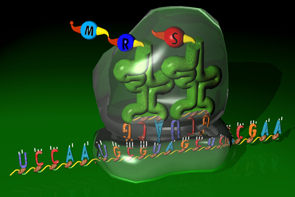
- Translation
- Happens in ribosomes in the cytoplasm (ribosomes are made of two halves, a small bottom half and a larger top half)
- mRNA takes copy of DNA template to cytoplasmic ribosomes
- tRNA brings amino acids to ribosomes where polypeptide synthesis occurs
- mRNA codons (sequence code) direct the sequence which tRNA amino acids come to the ribosome; results in particular order of amino acids in protein
- three RNA bases = one amino acid code

(found at http://www.coolschool.ca/lor/BI12/unit6/U06L01.htm)
- Three steps
- Initiation – mRNA binds to smaller ribosome half and is then joined by larger ribosome half
- Elongation – polypeptide lengthens one amino acid at a time
- Termination – occurs when RNA codon meaning “stop” shows up in ribosome’s system
- Result – one new protein for the cell’s metabolism

(found at http://genome.imim.es/courses/Madrid04/exercises/ensembl/images/ribosome.jpg)
- Here is a very simplified image of what transcription and translation are for:

(found at http://www.phschool.com/science/biology_place/biocoach/images/translation/centdogtl.gif)
- Regulated gene expression – determines which regions of DNA/genes are transcribed and translated; also how cell metabolism is controlled
- Four ways of regulated gene expression
- Transcriptional control – in nucleus; regulates which genes are transcribed and/or the transcription rate
- Posttranscriptional control – in nucleus; controls how and when mRNA is processed before heading into the cytoplasm
- Translational control – in cytoplasm, after mRNA leaves nucleus and before there is protein production; controls mRNA’s ability to bind ribosomes
- Posttranslational control – in cytoplasm, after protein synthesis; changes protein in order to make in functional
- When gene expression is not regulated, it leads to cancer
- Summary of above section – DNA makes RNA, which makes protein, which helps run cell metabolism
- This will probably be the century of biological technology, or DNA technology
Cancer – “example of what happens when genetic control goes awry” (found in slide 27 of Genetics – BIO 156 PowerPoint presentation)
- Cancer cells’ characteristics
- Cell looks abnormal and stops becoming the cell it’s supposed to be
- Abnormal chromosomes (in size and number), as well as abnormal nuclei
- Extra gene copies are often found
- Unlimited dividing ability – cancer cells essentially immortal, as they do not respond to inhibitory growth factors and also divide without being signaled by progressive growth factors
- Fails to go under apoptosis, programmed cell death
- Tumors happen when cells do not stop replicating when they should
- Tumors form – benign encapsulated and do not invade surrounding space; cancerous not held within boundaries and spreads to other body parts
- Go through angiogenesis and metastasis
- Angiogenesis – formation of new blood vessels
- Metastasis – when cancer cells begin new tumors far from the primary tumor; recovery doubtful in this stage
- Cell’s metabolism no longer under control
- Three phases of cancer cells
- Initiation – mutation of single cell that causes continual division
- Promotion – tumor develops and tumor cells mutate
- Progression – cell mutates so it can invade surrounding tissue; no longer isolated
- Types of cancer
- Carcinomas – of epithelial tissue – skin, breast, liver, pancreas, intestines, lungs, prostate, thyroid
- Adenocarcinomas – of glandular epithelial cells
- Sarcomas – of muscle and connective tissues – bones and fibrous tissues
- Leukemias – of the blood
- Lymphoma – of lymphatic tissue
- Lung cancer most common overall
- One in three Americans will deal with cancer
- Prognosis dependent on – whether tumor has spread to surrounding tissues and whether metastatic tumors are in distant body parts
- Causes of cancer – genetic and environmental
- Genetic
- Mutated copies of same gene from both parents can predispose person, e.g. genes BRCA1 and 2 (breast cancer), RB (eye tumor)
- Only one mutated copy of RET (thyroid cancer) gene needed for predisposition
- Environmental
- Ionizing radiation, e.g. UV light, too many X-rays, radon gas, nuke bombs
- Tobacco (smoke) – related to 80% of all cancers, more when combined with alcohol
- Pollutants – metals, dust, chemicals, pesticides, asbestos, etc.
- Viruses – parts of loose DNA that can trigger cancer
- Four DNA virus types – Hepatitis B (liver cancer), Hepatitis C (liver cancer), Epstein-Barr (Burkitt’s lymphoma), human papillomavirus (cervical cancer, can be caught in time with a pap smear)
- Seven cancer warning signs -- CAUTION
- Change in bowel or bladder habits
- A sore that does not heal
- Unusual bleeding or discharge
- Thickening or lump in breast or elsewhere
- Indigestion or difficulty in swallowing
- Obvious change in wart or mole
- Nagging cough or hoarseness
- These signs can be caused by something else, but a medical professional should be consulted nonetheless.
- Routine cancer screening tests
- Self-examination – monthly exams of breasts/testicles beginning at 20 years old; ABCD’s of skin cancer detection (see below)
- Colonoscopy – starting at 50 years old, every five years
- Mammogram – after 40 years old, yearly
- Pap smear – begin three years after sexual intercourse begins and/or no later than age 21
- Skin cancer detection ABCD’s
- A – asymmetry
- B – border is irregular
- C – color varies from one area to another
- D – diameter is larger than 6 mm
- These should not cause alarm, but should be looked at if present.
- Other forms of detecting cancer
- CAT scan
- MRI – useful for tumors in tissue surrounded by bone
- Ultrasound – useful for tumors in stomach, prostate, pancreas, kidney, uterus, ovary
- Tumor marker tests – tests blood for antigens/antibodies (e.g. colon, prostate, liver cancers)
- Genetic tests – “tests for mutations in proto-oncogenes and tumor-suppressor genes” (from slide 40 of Genetics – BIO 156 PowerPoint presentation)
- Needle biopsy can confirm if a tumor is malignant
- Standard cancer treatments
- Surgery – often preceded by and followed up with radiation therapy
- Radiation therapy – X-rays or gamma rays that are localized to disrupt cancer cell’s cycle; temporary side effects are – diahhrea, dry/red/irritated skin, dry mouth, fatigue/weakness, nausea, possibly permanent hair loss at site of treatment
- Chemotherapy – drugs for the whole body that kill cells by interfering with their DNA; can fail from cancer developing drug resistance
- Bone marrow transplants – take bone marrow from one person to another; sometimes done in conjunction with chemo
- Newer cancer therapies
- Immunotherapy – “inject immune cells that are genetically engineered to bear the tumor’s antigens” (from slide 41 of Genetics – BIO 156 PowerPoint presentation); immune cells produce cytokines which produce antigen to cytotoxic T cells, which in turn destroy tumor cells
- Passive immunotherapy – injections linked with radioactive isotopes or chemotherapeutic drugs are administered
- P53 gene therapy – “a retrovirus in clinical trial that is injected into the body where it will infect and kill only tumor cells (cells that lack p53 = tumor cells)” (from slide 42 of Genetics – BIO 156 PowerPoint presentation)
- Angiogenesis inhibition -- drugs called angiostatin and endostatin that inhibit angiogenesis are used
Meiosis and Inheritance – how genes are passed from parents to offspring
- Meiosis – two cell divisions turn out four haploid cells (one chromosome per cell instead of two)
- Meiosis occurs only in gametes (egg and sperm) – part of sexual reproduction
- Meiosis is part of spermatogenesis and oogenesis
- Crossing-over occurs between different pairs of chromosomes
- As a result, 8,388,608 different possible chromosome combinations are possible when beginning meiosis. This guarantees diversity in offspring.
- Chromosomes duplicate (as they do for mitosis) before meiosis 1
- Meiosis 1 – after first separation (meiosis), the two daughter cells still have two chromosomes each (like mitosis)
- Meiosis 2 – all four “granddaughter” cells have only one chromosome each, as they do not replicate before meiosis 2
- As a result of half the number of chromosomes in a finished meiotic cell than in a finished mitotic cell, when two meiotic cells fertilize (as meiosis is part of sexual reproduction), the product is an offspring with a full number of chromosomes, half from the mother and half from the father.
- Here is an image of meiosis

- Here is an image of the comparison of meiosis and mitosis

(found at http://anatomy.iupui.edu/courses/histo_D502/D502f04/lecture.f04/cell.f04/cellimages/Alb207.jpg)
- Genotype and Phenotype
- Genotype – genes of the individual
- Phenotype – physical characteristics resulting from genotype
- Alleles – “A particular gene, or protein-coding region of DNA along a chromosome might have a few different variations, called alleles” (from slide 47 of Genetics – BIO 156 PowerPoint presentation)
- Alleles divided into two categories – recessive and dominant
- Recessive notated by a lower-case letter
- Dominant notated by an upper-case letter
- Each pair of autosomes (non-sex chromosomes) carries alleles for particular traits
- Pairs are notated by letters, e.g. EE, Ee, ee
- EE – homozygous dominant genotype – shows dominant phenotype
- Ee – heterozygous genotype – shows dominant phenotype
- ee – homozygous recessive genotype – shows recessive phenotype
- Punnett’s square helps determine the outcome of genotypes and phenotypes in offspring according to the parent’s genotype pairs (e.g. EE).

(found at http://www.uic.edu/classes/bms/bms655/gfx/punnettsquare.gif)
§ Punnett’s square can also be used for two-trait crosses

(found at http://w1.avis.ne.jp/~hirocafe/columns/image/mendel_ChfaGrgr.gif)
- Combination of alleles in particular genes or chromosome regions in your parents determines hereditary traits, e.g. eye color
- Such alleles, or similarities in physical appearance based on genes and chromosomes, are evidenced in my second online lab, with both the dragon lab and the fly lab.
- Polygenic traits are controlled by several sets of alleles
- Diseases, as well as features, can be genetically coded and inherited. Sickle cell anemia is one such disease.
- Autosomal recessive disorders – Tay-Sachs, Cystic Fibrosis, PKU (Phenylketonuria), Sickle-Cell disease
- Autosomal dominant disorders – Marfan Syndrome,
- Fertilization
- Happens after meiosis when a male and female gametes join to form a new cell called a zygote, which, as I said earlier as well, contains all 23 chromosome pairs
- Zygote goes through mitosis while making its way through the fallopian tube
- After sufficient mitosis, zygote becomes morula when it reaches the uterus
- Normal development ensues as long as there are only two types of each chromosome.
- Abnormal chromosome sets – if there are three instead of two of a kind, it is called trisomy
- Most common trisomy is Down’s syndrome – trisome of chromosome 21

No comments:
Post a Comment