Cell Project
I began with these materials: four poster boards, two white and two lime green; an intermediate level K’Nex set; Gummy Rings; cherry tomatoes; a peach; Toffifay; brown rice (which ended up never being used); a bunch of Play-Doh; Sour Neon Worms; Gummy Worms; two balls of yarn, one red and white and the other green and white; “Beef Lit’l Smokies” mini hot dogs; some chocolate covered malt balls; and some yogurt covered raisins.


I started with making my basic cell model.
I started by making my plasma membrane (represented by blue Play-Doh).
Then, I added a flagella (green yarn), some lysosomes (Toffifays), and a couple of vesicles (chocolate malt balls; one undergoing exocytosis, and the other coming out of endocytosis).

Next, I added some mitochondria (mini hot dogs) and a Golgi apparatus (gummy rings), with “Golgi-looking” vesicles in the process of breaking off the Golgi apparatus.
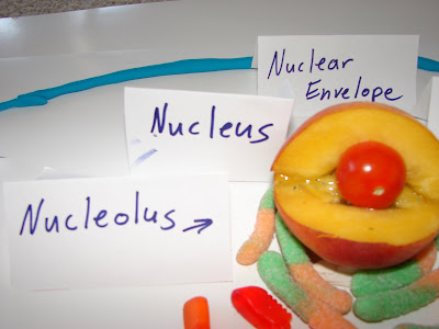
Lastly, I put in a nucleus (including nuclear envelope [peach fuzz/skin], nucleolus (cherry tomato), and chromatin [peach meat]), smooth and rough (sour worms with salt/sugar for ribosomes on rough, regular gummy worms for smooth) endoplasmic reticulum, a centriole (light lime-colored Play-Doh) and several ribosomes free in the cytoplasm (yogurt covered raisins).
Now the labels came out! Here is a series of pictures showing an overview of my cell with its labels, along with close-ups of each label and what it is labeling.
Vesicles after endocytosis are used to carry in useful objects from outside the cell into the inside of the cell.

Vesicles that go through exocytosis transport waste from inside the cell to outside the cell.

The nucleolus produces ribosomal RNA, which in turn joins with proteins in the nucleolus to form subunits of ribosomes.

The nucleus holds the DNA and RNA that hold and transfer genetic information vital to a cell’s metabolism, e.g. protein synthesis information.
The nuclear envelope allows useful objects in and out of the nucleus, e.g. mRNA out of nucleus.

The nucleus contains chromatin, which coils into chromosomes before the cell splits.
The rough ER makes protein with its ribosomes.
The smooth ER makes carbohydrates and lipids.
The Golgi apparatus packages proteins and lipids from the ERs to distribute around and outside the cell.
Mitochondria produce energy by cellular respiration.
Centrioles are made up of microtubules.

Ribosomes synthesize protein.

Lysosomes are made by the Golgi apparatus and transport Golgi’s proteins and lipids throughout the cell.

The flagella, which is made up of microtubules as well, helps particular cells (like sperm) move throughout the body.

Lastly, the plasma membrane transports molecules in and out of the cell, as well as separates the inside of the cell from the rest of the surrounding area.

Next is DNA replication, along with mitosis.
First, I made a K’Nex model of the double helix DNA.
Here is a close-up of the base pairs that make up the rungs of the DNA ladder.
Now, here’s a model of DNA replication.
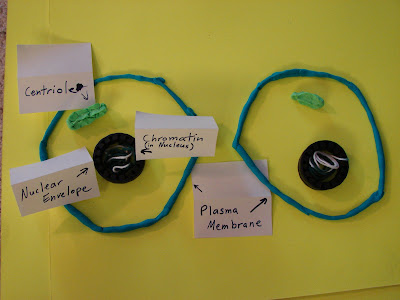
Now for mitosis. Here is a picture of prophase, when the chromosomes are beginning to condense in the nucleus.
Now for metaphase. This is after the DNA has replicated and the duplicated chromosomes are lined up down the center of the cell.

The next phase is anaphase. Here the chromosomes have split and are being drawn towards the centrioles at the poles by the spindles.
Lastly, there’s telophase. Here the plasma membrane begins to form a cleavage furrow on the way to splitting.
The end result is two daughter cells which develop their own separate nuclei in which the chromosomes turn back into indistinct chromatin.
Here is RNA, along with gene transcription and translation.
The first image is of mRNA copying a particular genetic protein pattern off of the DNA (also known as transcription). The DNA has the green and white backbones, while the mRNA has the yellow and white backbone.

After this transcription, the mRNA leaves the nucleus and attaches onto a cytoplasmic ribosome. Here, the mRNA has a three-base pair coding system (called codons) to signal the tRNA as to which amino acids to use. Meanwhile, the tRNA has picked up an amino acid from the cytoplasm and attached itself onto the ribosome above the mRNA. The tRNA also has an anti-codon to communicate with the mRNA as to which amino acids to use.

After this, the mRNA keeps running through the ribosome, as well as tRNAs moving in and out (from right to left) with fresh amino acids to pair up with the mRNA. After this happens several times, an amino acid chain is formed (called a polypeptide, which will eventually be a synthesized protein).

Finally, the mRNA sends through a signal to stop piling up the amino acids, and the whole system breaks apart, until it is time for the next protein to be synthesized. The now-free polypeptide is the newly synthesized protein resulting from this translation.

Conclusion
My project portrays several things. First, it illustrates the basic model of a cell, along with a short description of what each part was made from and how each part contributes to cell metabolism. Next, it portrays DNA and its replication with some K’Nex, as well as models of the stages of mitosis, also portrayed with some K’Nex and some Play-Doh. Lastly, it focuses on RNA and how it performs the transcription and translation of DNA information, resulting in protein synthesis.
I learned quite a few things from doing this project. First of all, I was reminded of how much work is involved in such endeavors. However, I was also reminded of the great value that results from doing a project like this. Because of this project, the basics of cell and genetic form and function were physically reinforced in my mind. As a result, I believe I will remember all this information better than if I had not done this project. Also, I learned how to organize the loads of information that I have been trying to digest over the past two weeks. Lastly, I learned how to enjoy this homework while working hard.


No comments:
Post a Comment